
Image
|
Figure Caption
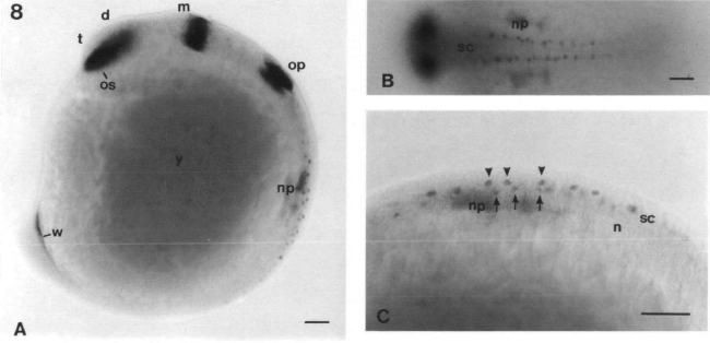
Fig. 8 In situ hybridization of pax[zf-b] on whole-mount embryos at 15 h of development. The embryo is oriented as in Fig. 3. (A) Lateral view of the embryo. (B) Dorsal view of the spinal cord of the embryo. (C) Close up of the spinal cord seen from lateral. Dorsal is to the top, the two rows of cells described in Discussion are marked with arrows and arrowheads, respectively. Abbreviations: d, diencephalon; e, eye; h, hindbrain; m, midbrain; np, nephritic primordium; os, optic stalk; ov, otic vesicle; sc, spinal cord; t, telencephalon; w, Wolffian duct; y, yolk. Bars: 50μm.
Figure Data
Acknowledgments
This image is the copyrighted work of the attributed author or publisher, and
ZFIN has permission only to display this image to its users.
Additional permissions should be obtained from the applicable author or publisher of the image.
Full text @ Development