
|
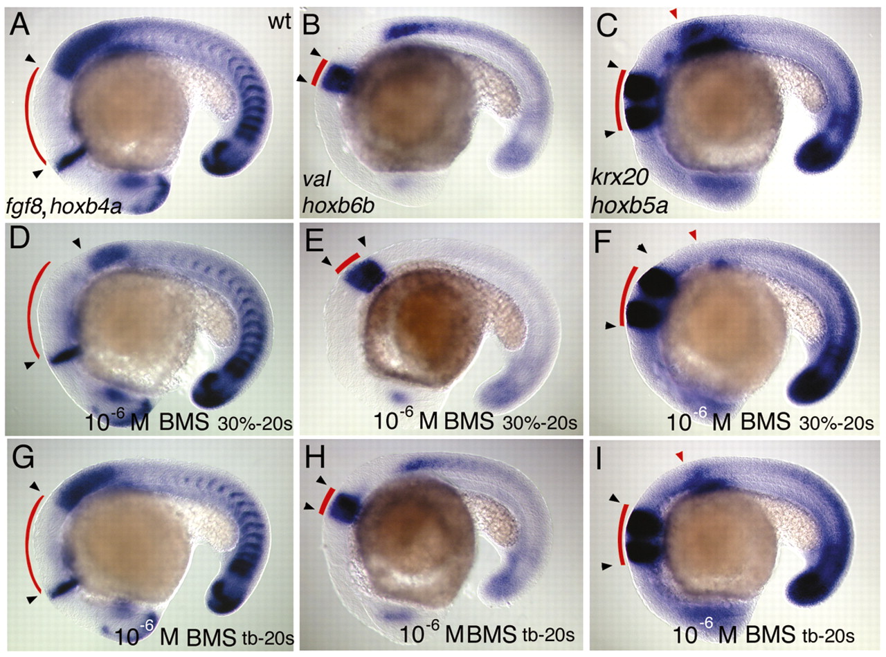
Fig. 7 In situ hybridisations at the 20s stage to visualise the effects of treating wild-type embryos with 10–6 M BMS493 during different time windows on the hindbrain and the spinal cord. All panels are lateral views, anterior is to the left. Curved red lines indicate the lengths of hindbrain segments of untreated wild types in all panels. Black arrowheads indicate the true extend of hindbrain segments observed in untreated wild-type (A-C) and experimental embryos. (A,D,G) Expression of fgf8 and hoxb4a; (B,E,H) val and hoxb6b and (C,F,I) krx20 and hoxb5a. (D,E,F) Inhibition of RA signalling by BMS493 from 30% epiboly onwards expands the hindbrain between the fgf8 and hoxb4a domains, as well as val and krx20 domains; (D,E,F) hoxb4a, hoxb6b and the anterior part of the hoxb5a domain (red arrowhead) are strongly downregulated in the spinal cord. (G,H,I) BMS493 treatments that exclude pre-segmentation stages neither lead to expansion of the hindbrain nor to strong reduction of hoxb4a, hoxb6b and hoxb5a.