
|
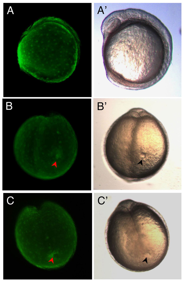
Fig. S4 Targeting of eGFP mRNA to DFCs and expression in Kupffer’s Vesicle. (A-C, A′-C′) Live embryos at 7-somite stage. (A-C) Fluorescence images of eGFPDFCOE embryos showing eGFP expression only in the yolk and KV and not in the rest of the embryo. (A′-C′) Corresponding transmitted light images of embryos in A-C. (B) Occasionally, eGFP could be found filling the entire KV (red arrowhead), whereas in the majority of embryos, such as in (C), eGFP only outlines part of the KV (red arrowhead), implying that only a subset of DFCs take up and/or express eGFP RNA/protein. Embryos in (B′ and C′) showing fully formed KV (black arrowheads). All eGFPDFCOE embryos (n=160) developed normally. (A and A′) Lateral view. (B, B′, C, and C′) Posterior views.