
|
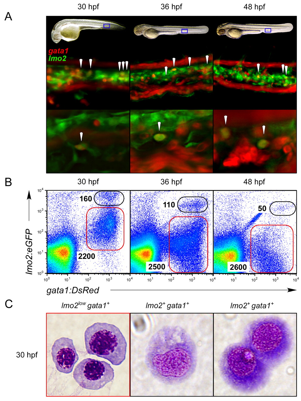
Fig. 2 Coexpression of lmo2 and gata1 reveals immature hematopoietic precursors in the PBI. (A) Fluorescence microscopy reveals cells within the vascular plexus of the PBI that expressed both gata1:DsRed and lmo2:eGFP fluorophores (arrowheads) in double transgenic animals. Blue boxes in embryonic images in upper panels denote the regions shown at 200x magnification in middle and 400x magnification in lower panels at 30, 36 and 48 hpf. Lower panels show a single, deconvolved z slice, demonstrating coexpression of each transgene in single cells (arrowheads). (B) Cells coexpressing the gata1:DsRed and lmo2:eGFP transgenes prospectively isolated by flow cytometry. Double-positive cells peak in number at 30 hpf, with approximately 160 cells per embryo (left panel). (C) Compared with purified primitive erythroblasts sorted by low levels of lmo2:eGFP and high levels of gata1:DsRed (red gate in 30 hpf plot in B), cytological staining of purified 30 hpf lmo2+ gata1+ cells (black gate) showed immature morphologies indicative of early hematopoietic progenitors. Magnification, x1000.