
|
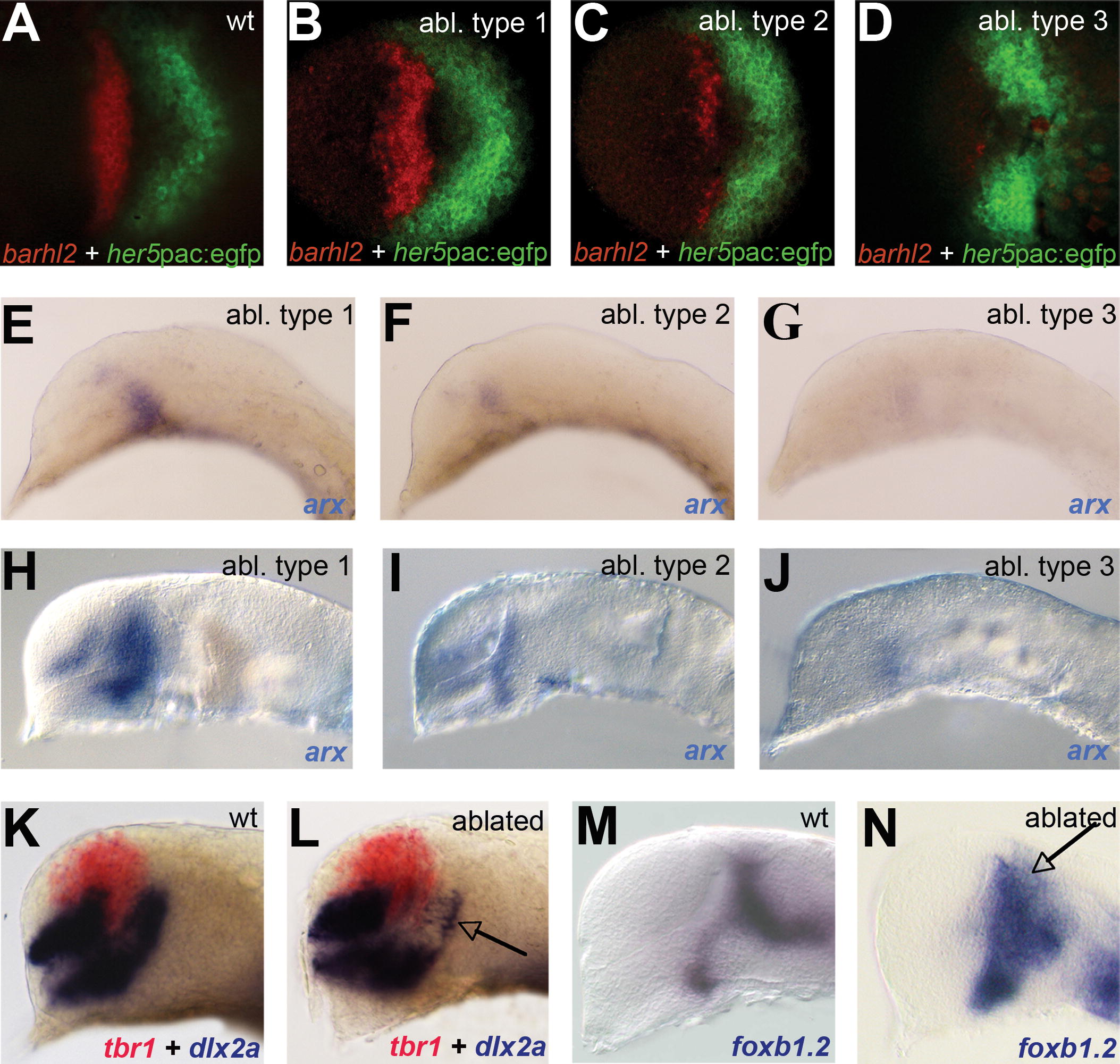
Fig. 6 Ablation of Prethalamic Precursors (A–D) barhl2 (red) and her5pac:egfp (green) are shown in (A) wild-type (wt) and (B–D) prethalamus-ablated (abl) embryos 2 h after surgery. (E–G) arx expression at the 12-somite stage and (H–J) prim5 stage in ablated embryos are shown, representing the different ablation types. (K and L) tbr1 is shown in red, and dlx2a in blue; in comparison to the wt (K), the prethalamic expression of dlx2a is reduced in ablated embryos (arrow in [L]). (M and N) foxb1.2 expression at prim 5 shows that mid-diencephalic domains in ablated embryos are not impaired although the expression appears unorganized in the thalamic domain (arrow in [N]).