
|
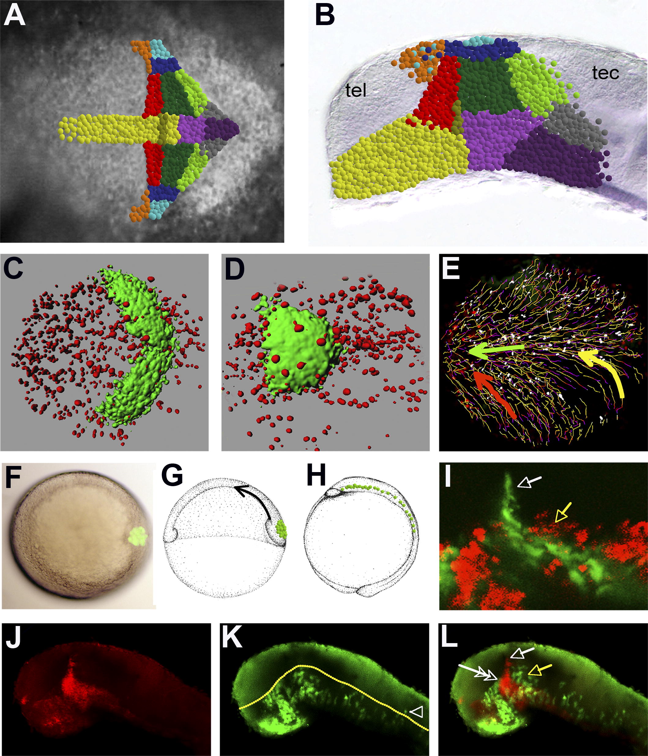
Fig. 5 Cell Movement in the Neural Plate (A and B) Schematic overview of the results obtained by the fate-mapping experiments. Colours at prim5 stage correspond to the territories labelled at bud stage. (C–E) Time-lapse analysis of nuclei (red) movement in the neural plate in her5pac:egfp (green): 3D rendering of a 200-μm z-series done at (C) bud stage and (D) 5-somite stage. (E) Schematic overview of the trajectories of individual nuclei over the recorded time (time-lapse movie can be seen in Video S1), with labelled nuclei in white. Wavy lines and dark to light colours of the lines represent the timeline of nuclei movement. Some lines are shorter because nuclei moved out of or into the observed area over the recorded time. We observed three types of movement: the basal cells move anteriorly (green arrow), posterior alar cells move towards the midline and then anteriorly (yellow arrow), and anterior alar cells move diagonally towards the midline (red arrow). (F–L) Transplantation of basal cells: (F) animal pole view in which labelled cells (green) are transplanted on top of the shield; (G) schematic lateral view of shield stage embryo in which transplanted cells are going to move towards the animal pole during gastrulation; (H) schematic lateral view of bud stage embryo in which transplanted cells are spread along the midline of the embryo; and (I) shhGFP is shown in green, transplanted cells in red, at 22 hpf in a live embryo. (J–L) shh is shown in red, transplanted cells in green; at 30 hpf, basally derived cells form a large proportion of the brain, showing that just the tip of the ZLI is formed by alar plate cells (white arrow), the basal ZLI is composed of basal cells (white double arrow), and the ventral part of the developing thalamus is built by basal cells (yellow arrow). The yellow line in (K) indicates the border between alar and basal plate; the arrowhead in (K) points to a single basal cell moving alar.