
|
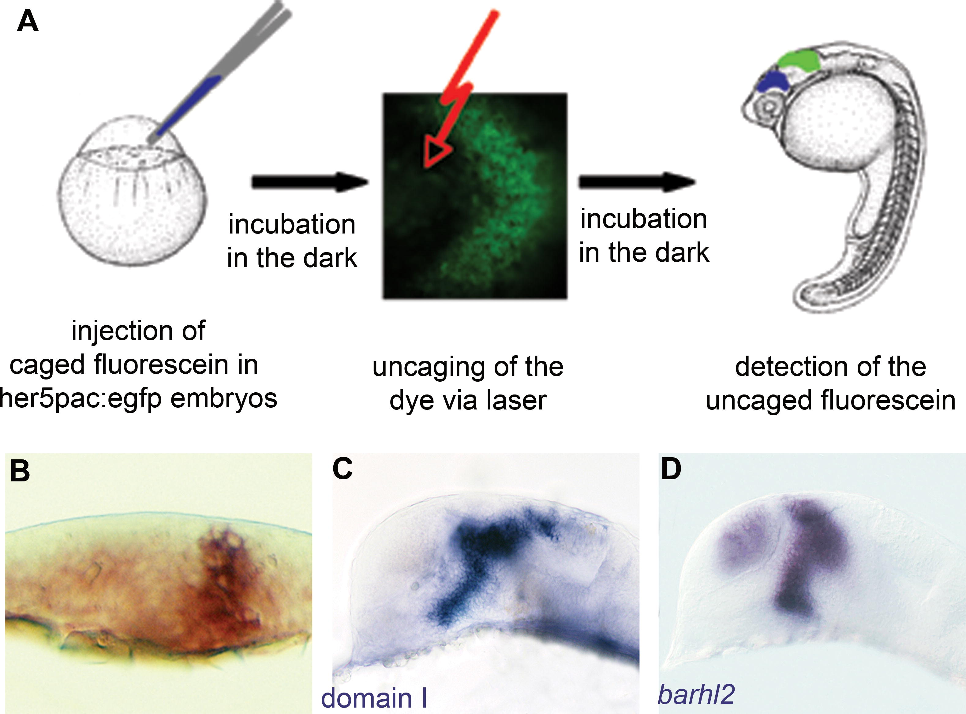
Fig. 3 Schematic of the Uncaging Procedure (A) Caged fluorescein is injected into one-cell stage her5pac:egfp embryos. Injected embryos are kept in the dark until the fluorescein is uncaged in 6–10 cells of the diencephalic neural plate at bud stage using a laser beam. After further light-protected incubation, the embryos are fixed at prim5 stage, and the uncaged form of the fluorescein is detected via antibody staining. (B) Transverse section of a neural plate after the uncaging experiment, cells of the whole z-axis are labelled. (C) Result of labelling cells via uncaging in domain I, which correlates mostly with the expression pattern of barhl2 at bud stage. (D) At prim5, labelled areas in the diencephalon resemble the endogenous expression pattern of barhl2.