
Image
|
Figure Caption
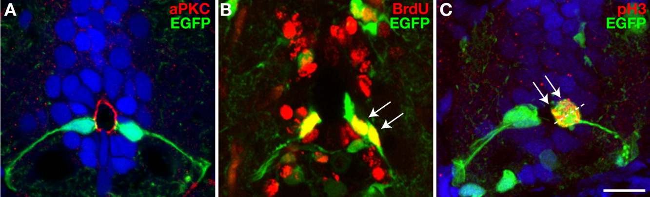
Fig. 4 olig2+ radial glial cells have asymmetric features. A-C: Transverse sections of 15-dpf Tg(olig2:egfp) fish, dorsal up. A: aPKC (red) was localized to the apical membranes of EGFP+ radial glia (green) at the central canal. B: Two BrdU+ EGFP+ cells (arrows) were next to each other near the central canal. These cells might have been recently divided siblings. C: Anti-phospho-Histone H3 (pH3) antibody labeling revealed chromosomes (arrows) aligned with the axis of division perpendicular to the central canal (dashed line). Scale bar = 10 μM.
Figure Data
Acknowledgments
This image is the copyrighted work of the attributed author or publisher, and
ZFIN has permission only to display this image to its users.
Additional permissions should be obtained from the applicable author or publisher of the image.
Full text @ Dev. Dyn.