
|
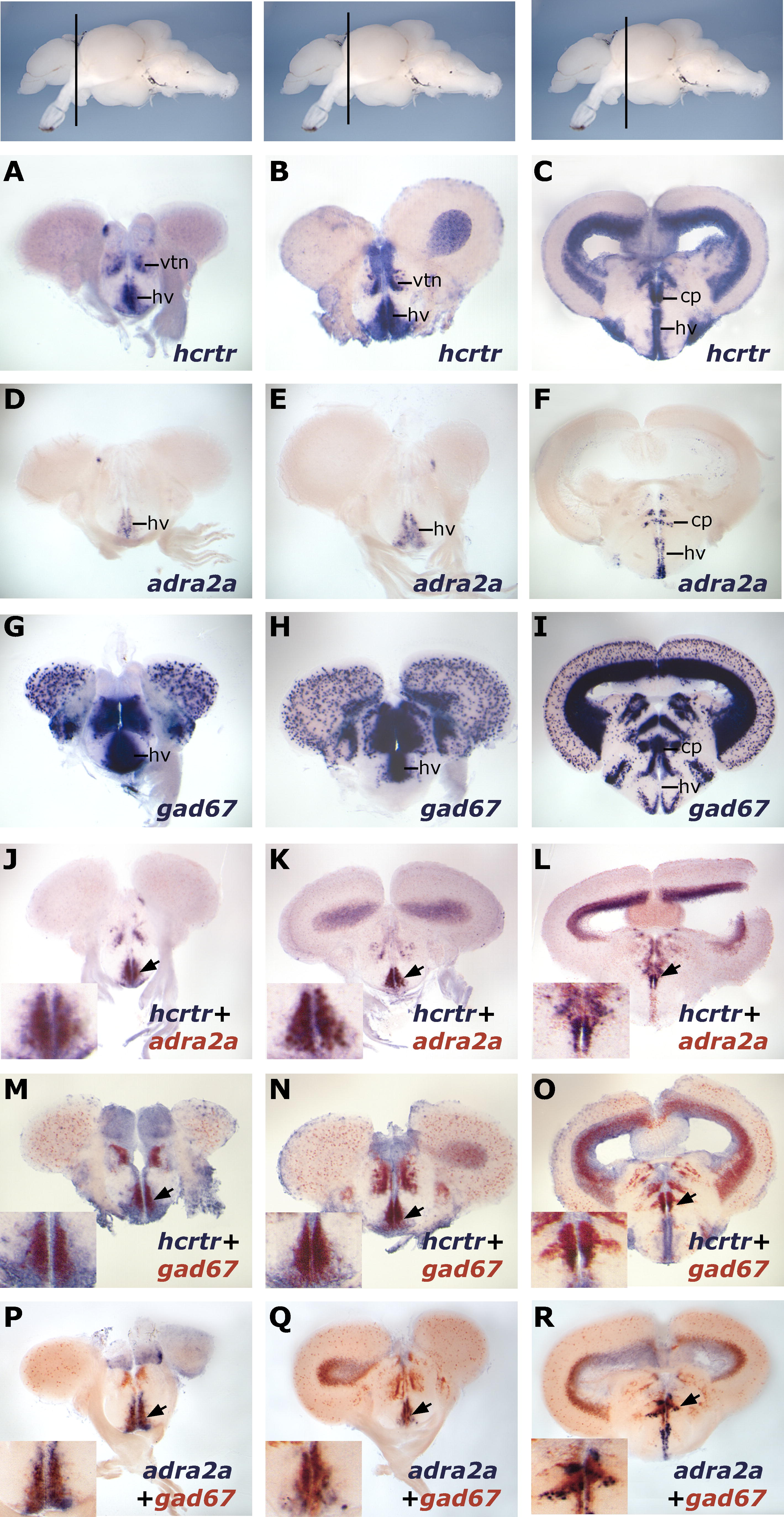
Fig. 6 A Population of Anterior Hypothalamic hcrtr-Positive Neurons is adra2a- and gad67-Positive. The topmost panels display lateral views of zebrafish adult brain with transversal plane corresponding to sections presented below. Results were all obtained using ISH. (A–I) Single ISH expression patterns of hcrtr (A–C), adra2a (D–F), and gad67 (G–I) in the anterior diencephalon. In the most rostral part of the diencephalon, hcrtr is mainly expressed in the ventral thalamic nucleus (vtn) and the ventral zone of the periventricular hypothalamus (hv) (A and B). Posteriorly, it is mostly expressed in the peripheral gray zone and in periventricular areas including the central posterior thalamic nucleus (cp) and the ventral zone of the periventricular hypothalamus. (J–L) Double ISH showing colocalization of hcrtr and adra2a in the ventral zone of the periventricular hypothalamus (arrowheads). (M–O) Double ISH showing colocalization of hcrtr and gad67 in the ventral zone of the periventricular hypothalamus and ventral thalamic nucleus (arrowheads). (P–R) Double ISH showing colocalization of gad67 and adra2a in the ventral zone of the periventricular hypothalamus (arrowheads). Note that hcrtr, gad67, and adra2a only colocalize in the anterior hypothalamus and not in the posterior ventral zone of the periventricular hypothalamus, where gad67 is absent (I). Posterior to this area, only the central thalamic nucleus expresses these three markers (last column).