
|
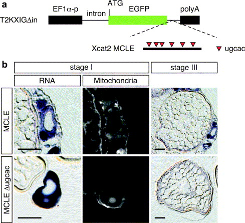
Fig. 3 The Xcat2 3′ UTR directs localization to the mitochondria cloud in transgenic zebrafish oocytes. (a) Schematic representation of the transgenic construct used to express a GFP mRNA fused with Xcat2 3′ UTR. Xcat2 MCLE or Xcat2 MCLEΔugcac 3′ UTR (Betley et al., 2002) was inserted into the downstream site of EGFP in the T2KXIGΔin plasmid vector (Kawakami et al., 2004). Expression of GFP mRNA is driven by the EF1α promoter. (b) In situ hybridization in transgenic zebrafish oocytes expressing GFP mRNA fused with Xcat2 MCLE (upper panel) or MCLEΔugcac (lower panel), using antisense GFP RNA as a probe. Left column, expression of the reporter mRNA in a stage I oocyte; middle column, the same oocyte counterstained with Mitotracker. Right column, expression of the reporter mRNA in a stage III oocyte. Sections were cut at a thickness of 10 μm. Scale bars, 50 μm.
Reprinted from Mechanisms of Development, 124(4), Kosaka, K., Kawakami, K., Sakamoto, H., and Inoue, K., Spatiotemporal localization of germ plasm RNAs during zebrafish oogenesis, 279-289, Copyright (2007) with permission from Elsevier. Full text @ Mech. Dev.