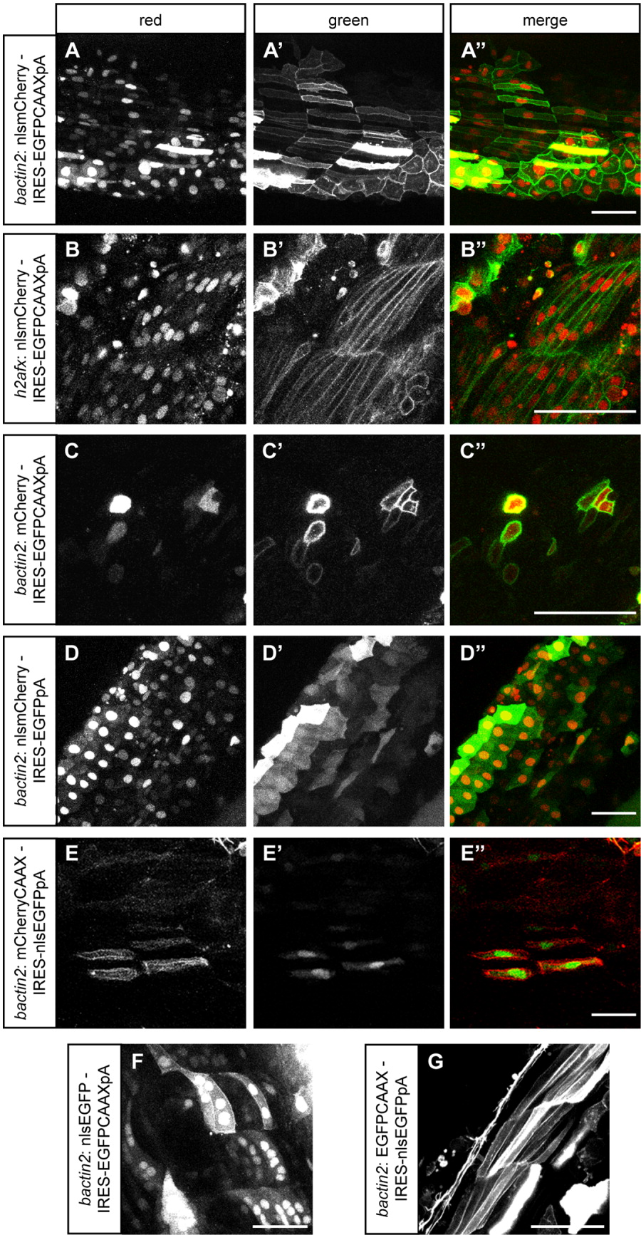
Fig. 2 Validation of reporters and internal ribosome entry sequence (IRES) constructs. Embryos shown were injected at the one-cell stage with expression constructs made with the pISce-Dest destination vector, then mounted at 24 hours postfertilization (hpf) for confocal microscopy of the trunk. All plasmids tested generate functional bicistronic messages, as demonstrated by the presence of both mCherry and EGFP fluorescence. A-A″:bactin2: nlsmCherry-IRES-EGFPCAAXpA. B-B″:h2afx: nlsmCherry-IRES-EGFPCAAXpA. C-C″:bactin2: mCherry-IRES-EGFPCAAXpA. D-D″:bactin2: nlsmCherry-IRES-EGFPpA. E-E″:bactin2: mCherryCAAX-IRES-nlsEGFPpA. F:bactin2: nlsEGFP-IRES-EGFPCAAXpA. G:bactin2: EGFPCAAX-IRES-nlsEGFPpA. Scale bar = 50 μm.
                
                    
                        Image


                        Figure Caption


                        Acknowledgments


This image is the copyrighted work of the attributed author or publisher, and
ZFIN has permission only to display this image to its users.
Additional permissions should be obtained from the applicable author or publisher of the image.
      Full text @ Dev. Dyn.