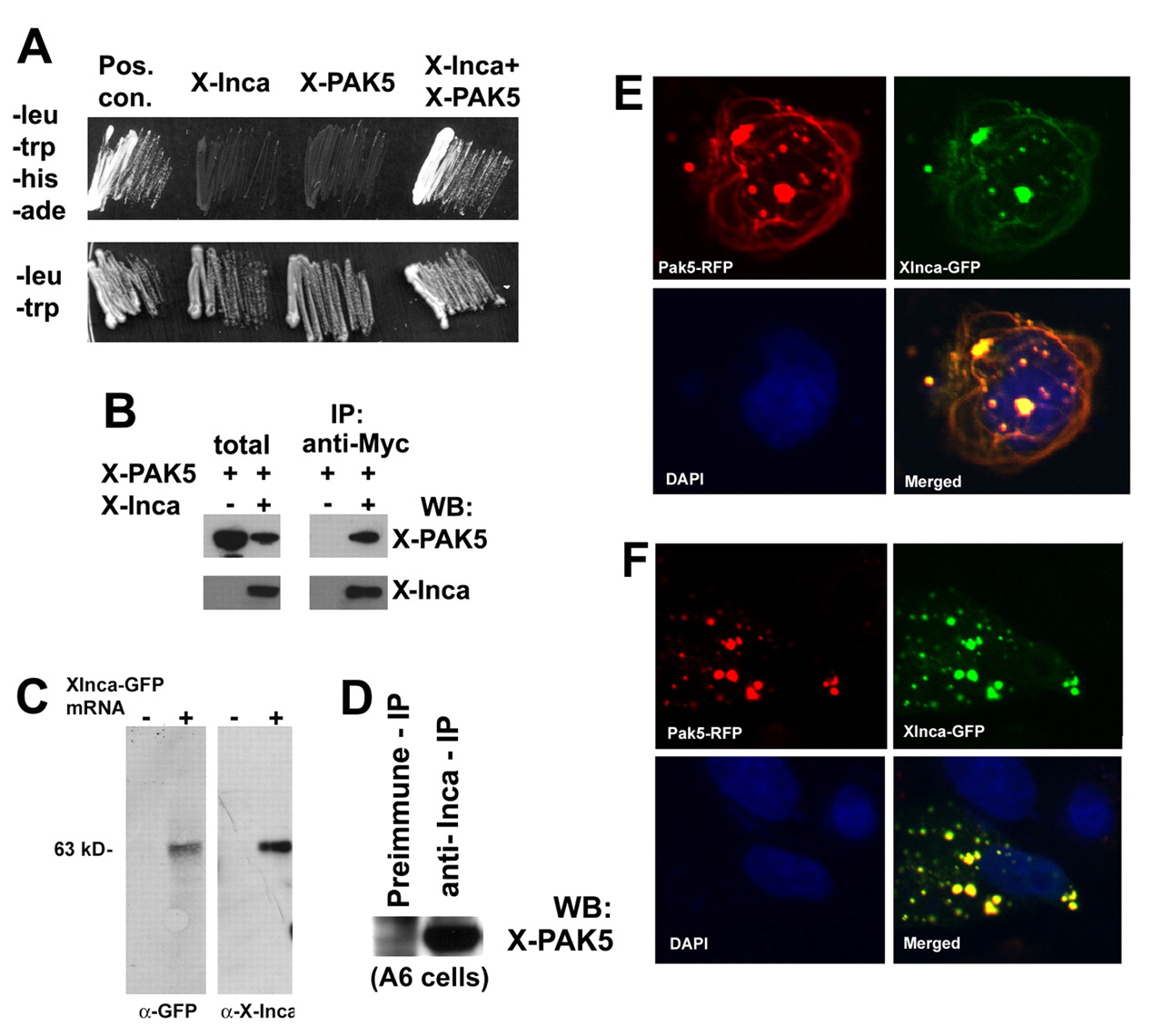
Fig. 6 Inca interacts with PAK5. (A) Selective medium streaks of yeast cells expressing Xenopus Inca and PAK5, either alone or together, as indicated. Inca+PAK5 allows growth on medium lacking leucine, tryptophan, histidine and adenine. Positive controls include transfection with vectors containing T antigen and p53 (Clontech). Streaks on -leu/-trp (lower panel) shown as a control for transfection. (B) Extracts prepared from HEK293 cells transfected with expression plasmids encoding a PAK5-GFP fusion or a Myc epitope-tagged Inca separately or together, immunoprecipitated with anti-Myc, followed by western blot with antibody for GFP or Myc. Lanes labeled as total are from lysates prior to immunoprecipitation. PAK5-GFP precipitates with the anti-Myc antibody, but only when Myc-tagged Inca is present in the extract. (C) Anti-IncaA peptide antibody specificity. Fertilized eggs were injected with 1 ng of synthetic mRNA encoding XInca-GFP, and cultured to stage 20. Detergent (1% NP40)-solubilized protein was extracted with 1,1,2-trichlorotrifluoroethane (Freon) to remove yolk and the equivalent of two embryos analyzed by SDS-PAGE/western blot. Both anti-GFP and anti-Inca recognized a single band of the correct molecular weight (∼63 kDa). (D) Extract from untransfected Xenopus A6 cells immunoprecipitated with preimmune serum or anti-Inca, followed by western blot using antiserum raised against PAK5 (residues 122-224, a generous gift of N. Morin). Immunoprecipitation with anti-Inca enriches the PAK5 signal several-fold compared with preimmune serum, indicating Inca-PAK5 interaction. (E) Fluorescent images of a CHO cell cotransfected with plasmids encoding a PAK5-RFP fusion and Xenopus Inca-GFP fusion showing extensive overlap. (F) Fibrous structures positive for Inca and PAK5 are nocodazole-sensitive. CHO cells transiently transfected with PAK5-RFP and XInca-GFP were treated for 1 hour with 3 ng/ml nocodazole (Sigma), then fixed with methanol and photographed using an inverted fluorescence microscope. The fibers visible without nocodazole treatment (E) have disappeared.
Image
Figure Caption
Acknowledgments
This image is the copyrighted work of the attributed author or publisher, and
ZFIN has permission only to display this image to its users.
Additional permissions should be obtained from the applicable author or publisher of the image.
Full text @ Development

