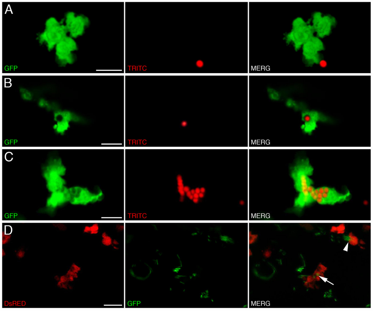
Fig. 7 Labeled cells within transgenic larvae display phagocytic activity. Phagocytosis of red fluorescent microspheres by labeled cells within 4 dpf lysC::EGFP larvae. (A-C) Summed Z stacks through individual EGFP-labeled cells within 4 dpf transgenic larvae approximately 6 hours following injection of fluorescent microspheres. (D) Summed Z stacks through posterior intestine of DsRED2-expressing cells containing ingested GFP-labeled Salmonella following infection of lysC::DsRED2 larvae at 5 dpf (images captured at 8 dpf). Arrow denotes labeled cell containing GFP-labeled bacteria, arrowhead denotes a labeled cell actively phagocytosing a microcolony of GFP-expressing bacteria. Scale bars: 10 μm.
                
                    
                        Image


                        Figure Caption


                        Acknowledgments


This image is the copyrighted work of the attributed author or publisher, and
ZFIN has permission only to display this image to its users.
Additional permissions should be obtained from the applicable author or publisher of the image.
      Full text @ BMC Dev. Biol.


        