
|
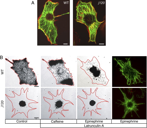
Fig. 2 Partial Dispersion in j120 Melanocytes Does Not Involve an Abnormal Microtubule or Actin Cytoskeleton and Is Independent of Actin (A) Fluorescent images of hodamine-phalloidin-labeled actin (red) and immuno-labeled α-tubulin (green) in cultured wild-type and j120 mutant melanocytes. Microtubule and actin cytoskeletons appear normal in the mutant. (B) Aggregation and dispersion of melanosomes in the absence of actin filaments. Melanocytes were treated with 5 μM latrunculin A for 15 min for disruption of actin filaments and then with caffeine for inducing dispersion. In latrunculin, wild-type melanocytes hyperdisperse their melanosomes, whereas the j120 mutant melanocyte still only partially dispersed its melanosomes. Aggregation (induced by epinephrine) is unaffected by latrunculin. Rhodamine-phalloidin staining confirms the loss of actin filaments in the latrunculin-treated melanocytes. Microtubules (green) were labeled with an antibody against α-tubulin. (Scale bars represent 10 μm.)