
|
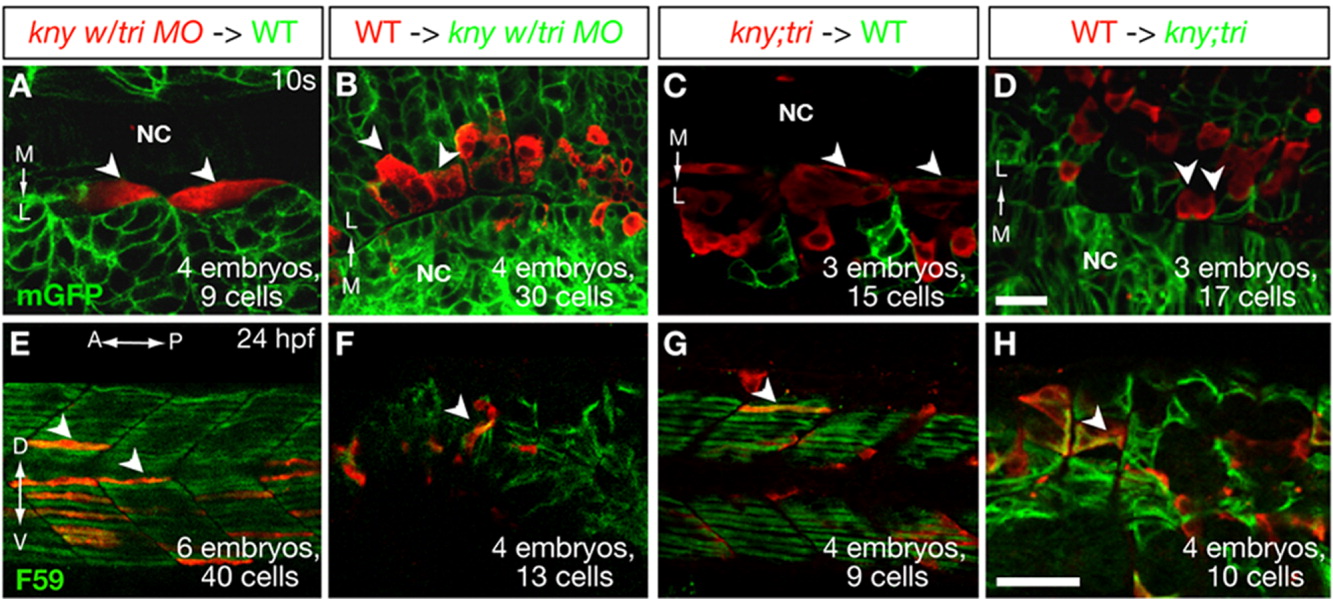
Fig. 3 Kny and Tri act cell non-autonomously during slow muscle morphogenesis. A-H: Confocal images of the host embryos expressing mGFP (A-D) or stained with F59 antibody (E-H). Transplanted donor cells are labeled with Rhodamine dextran (red). The white arrowheads highlight several representative donor cells. A, B, E, F: Transplantation experiments using kny embryos injected with tri MO as donors (A, E) or hosts (B, F). C, D, G, H: Transplantation analyses using kny;tri double mutants as donors (C, G) or hosts (D, H). A-D: Dorsal views of the host embryos at the 10-somite stage (14 hpf). E-H: Lateral views of the host embryos at 24 hpf. A, anterior; D, dorsal; P, posterior; L, lateral; M, medial; NC, notochord; V, ventral. Scale bars = 20 μm (A-D), 50 μm (E-H).