
|
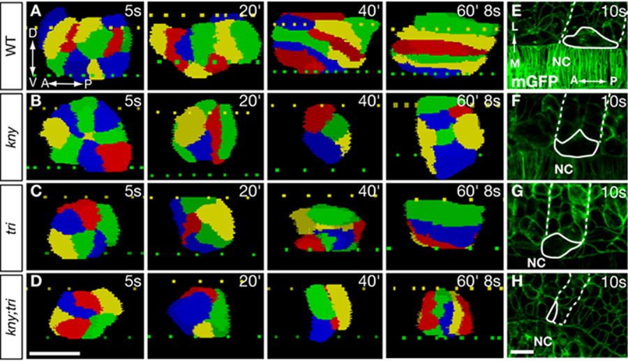
Fig. 2 The kny;tri adaxial cells fail to undergo proper shape changes that precede the lateral migration. A-D: 3D reconstruction of the adaxial cells within the third somite (see Experimental Procedures section). Lateral views. Individual cells are colored for clarity. Dashed lines denote the dorsal (yellow) and ventral (green) surfaces of the notochord. E-H: Dorsal views of the embryos expressing mGFP, one adaxial cell (solid line) and the corresponding somitic boundaries (dashed line) are highlighted. A, anterior; D, dorsal; L, lateral; M, medial; NC, notochord; P, posterior; V, ventral. Scale bars = 20 μm (A-H).