
|
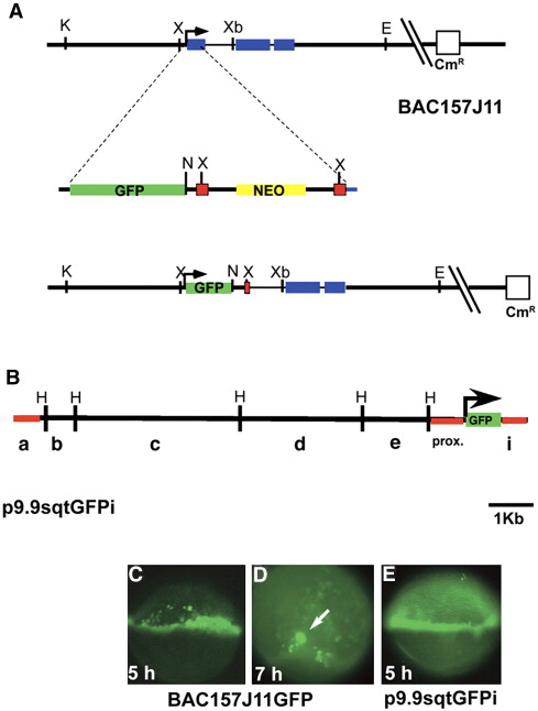
Fig. 1 Construction and expression of transient sqtGFP lines. (A) Diagram of the sqt locus in BAC157J11. sqt has three exons (blue rectangles) separated by two introns, 631 bp and 81 bp long, respectively. The start of transcription is indicated (arrow). Key restriction sites and the vector gene encoding chloramphenicol resistance are depicted. The GFP-FRT-neo-FRT cassette is diagrammed below the BAC, showing the site of integration in the first exon. The resulting engineered BAC clone is depicted below. After integration and excision of the gene encoding neomycin resistance, the gfp coding region replaces the sqt first exon. Green rectangle = GFP sequences; yellow rectangle = neomycin resistance gene; red squares = FRT recombination sites; blue rectangle = sqt coding sequences. (B) Diagram of p10sqtGFP following excision from the BAC by gap-repair. HindIII sites are indicated. Red lines indicate the sequences used to generate Tg-SqtapGFP and its derivatives. (C) Fluorescent image of a living, 5 hpf embryo injected with BAC157J11sqtGFP at the one-cell stage. Fluorescence appears throughout the margin, including the YSL, EVL and blastomeres. Perduring GFP expression is also observed in some cells farther from the margin. (D) Low magnification fluorescent image of a living, 7 hpf embryos injected with BAC157J11sqtGFP. Fluorescence is observed in the dorsal forerunners (white arrows). Perdurant expression is also detected in blastomeres and EVL cells farther from the margin. (E) Fluorescent image of a living 5 hpf embryo injected with p9.9sqtGFPi at the one-cell stage. GFP fluorescence is detected around the entire margin, in the blastomeres, EVL and YSL. K = KpnI; X = XhoI; Xb = XbaI; E = EcoR1; N = Not1; H = HindIII.
Reprinted from Developmental Biology, 310(2), Fan, X., Hagos, E.G., Xu, B., Sias, C., Kawakami, K., Burdine, R.D., and Dougan, S.T., Nodal signals mediate interactions between the extra-embryonic and embryonic tissues in zebrafish, 363-378, Copyright (2007) with permission from Elsevier. Full text @ Dev. Biol.