
|
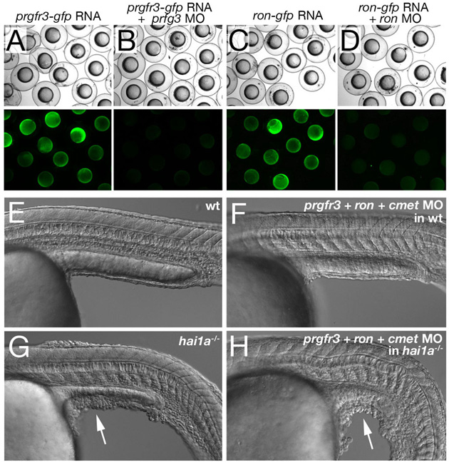
Fig. S2 Concomitant inactivation of cMet and its related receptors Prgfr3 and Ron is insufficient to rescue the epidermal defects of hai1a mutants. (A-D) prgfr3 and ron MOs efficiently block the translation of their respective mRNAs encoding Prgfr3-GFP or Ron-GFP fusion proteins. Upper panels show bright-field images, lower panel GFP fluorescence of embryos at the 80% epiboly stage, after injection of prgfr3-gfp mRNA (A,B) or ron-gfp mRNA (C,D), and with (B,D) or without (A,C) the respective MOs. Co-injection of MOs leads to complete suppression of GFP fluorescence. (E-F) Lateral Nomarski images of 24 hpf un-injected wild-type embryo (E) and wild-type embryos co-injected with cmet MO, prgfr3 MO and ron MO (each at 0.16 mM) (F), revealing that all three receptors are dispensable for normal skin development. (G,H) Lateral Nomarski images of 24 hpf un-injected hai1a mutant (G) and hai1a mutant co-injected with cmet MO, prgfr3 MO and ron MO (each at 0.16 mM), revealing that Hai1 does not act by inactivating cMet, Prgfr3 and Ron. White arrows point to dissociating epidermis on yolk extension.