
|
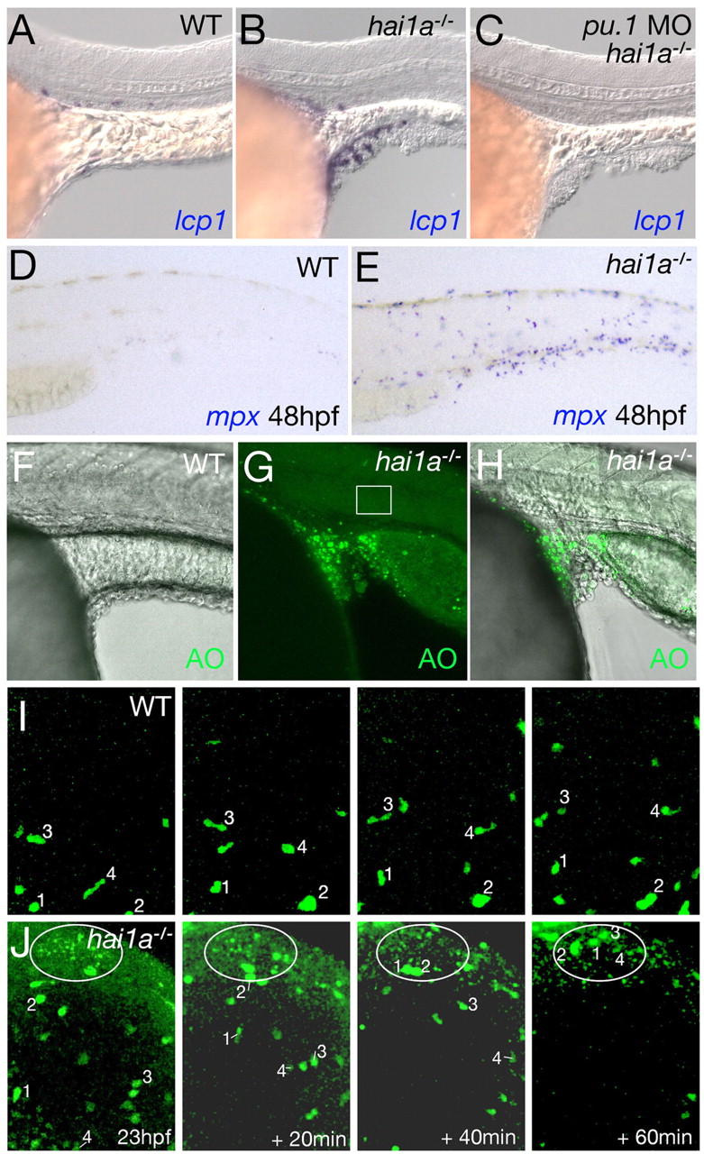
Fig. 6 Skin inflammation in hai1a mutants is linked to keratinocyte death, but is dispensable for defects in epithelial integrity. (A-C) Lateral views of yolk sac extension of 24 hpf embryos, after in situ hybridization for the leukocyte marker lcp1. Leukocytes accumulate around epidermal aggregates in hai1a mutants (B); this does not occur in wild-type siblings (WT; A). hai1a mutants injected with pu.1 MO lack leukocytes but display epidermal aggregates of equal severity (C). (D,E) Lateral trunk views of a wild-type sibling embryo (D) and a hai1a mutant (E) after in situ hybridization for the neutrophil marker mpx at 48 hpf. (F-H) Lateral views of yolk sac extension of a 24 hpf sibling embryo (F) and hai1a mutant (G,H), stained with acridine orange (AO). (F,H) Overlays of fluorescent and Nomarski images; (G) fluorescent image. Box in G marks the site where Movie 2 in the supplementary material was taken, stills of which are shown in Fig. 5B. (I,J) Stills from time-lapse Movies 6 and 7 in the supplementary material (at the indicated times after 23 hpf), demonstrating that Tg(fli1a:egfp)-marked leukocytes of hai1a mutants migrate from their site of origin anterior to the cardiac field over the yolk sac (Herbomel et al., 1999) to sites with nascent epidermal aggregates and numerous apoptotic cells (J). A cluster of acridine orange (AO)-positive cells is outlined. By comparison, leukocytes of wild-type siblings move more slowly and in a less directed fashion (I). For tracking, four individual leukocytes are indicated with numbers.