
|
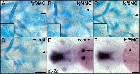
Fig. 8 Teeth develop relatively normally after morpholino antisense inhibition of fgf3 and fgf8, but subtle tooth effects are observed after fgf4 inhibition. (A–D) Ventral view of the pharyngeal region of morpholino (MO) injected, cartilage-stained specimens at 5 days with magnified teeth shown in inset. (A) Ceratobranchial cartilages were often completely missing after fgf3 MO injection, but teeth appeared normal (arrow). (B) After fgf4 MO injection, ceratobranchial cartilages were also reduced, and teeth, although always present, were often misshapen (arrow). (C) fgf8 morpholino injected fish display severe cartilage reductions, but teeth always developed normally (arrow). (D) Injection control displaying normal cartilages and teeth (arrow). (E and F) Dorsal views of control and fgf8 MO injected fish at 56 h. dlx2b expression in tooth germs (arrow, E) was variably affected: sometimes normal, sometimes missing, and sometimes reduced (arrow, F). Anterior is to the left in all panels. Scale bars = 100 μM.
Reprinted from Developmental Biology, 274(1), Jackman, W.R., Draper, B.W., and Stock, D.W., Fgf signaling is required for zebrafish tooth development, 139-157, Copyright (2004) with permission from Elsevier. Full text @ Dev. Biol.