
|
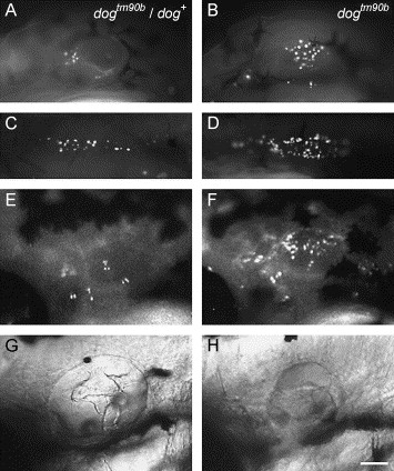
Fig. 7 Time course of cell death in dogtm90b. Single wild-type (A, C, E, and G) and dogtm90b mutant (B, D, F, and H) embryos stained with acridine orange at 48 hpf (A–D) and 72 hpf (E and F). (A and B) dogtm90b embryos show an increase in the number of acridine orange-positive cells in the otic vesicle (B) and migrating primordium of the posterior lateral line (D) relative to wild-type siblings (compare A, B and C, D). (E and F) The same wild-type sibling (E) and dogtm90b (F) embryos as shown in the previous panels continue to exhibit an increased number of AO-positive cells at 46 hpf. (G and H) Otocyst phenotype of dogtm90b (H) and wild-type sibling (G) embryos shown in (A, C, and E) and (B, D, and F), respectively. All panels: anterior to left, dorsal to top. Scale bar, 100μm.
Reprinted from Developmental Biology, 277(1), Kozlowski, D.J., Whitfield, T.T., Hukriede, N.A., Lam, W.K., and Weinberg, E.S., The zebrafish dog-eared mutation disrupts eya1, a gene required for cell survival and differentiation in the inner ear and lateral line, 27-41, Copyright (2005) with permission from Elsevier. Full text @ Dev. Biol.