
Image
|
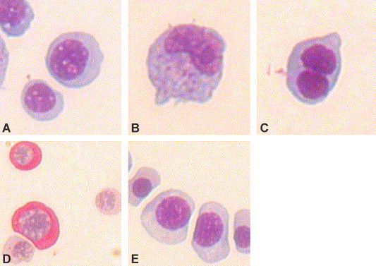
Figure Caption
Fig. 5 Wright staining of cytospin preparation of cells dissected out from the circulation of the ChdMO and wild-type embryos at 48 hpf. Most of the circulating cells are erythroid in morphology (A), monocytes (B), and binucleated erythroid cells (C) accounted for 1.2 and 1.4 per 200 cells, respectively. Periodic Acid Schiff (PAS) staining showed positivity in 21% of cells (D). (E) Circulating erythroid cells in the WT embryos. 2.0 per 200 cells in the WT embryos were binucleated (not shown). Monocytes were not evident. PAS staining was all negative in WT embryos.
Acknowledgments
This image is the copyrighted work of the attributed author or publisher, and
ZFIN has permission only to display this image to its users.
Additional permissions should be obtained from the applicable author or publisher of the image.
Reprinted from Developmental Biology, 277(1), Leung, A.Y., Mendenhall, E.M., Kwan, T.T., Liang, R., Eckfeldt, C., Chen, E., Hammerschmidt, M., Grindley, S., Ekker, S.C., and Verfaillie, C.M., Characterization of expanded intermediate cell mass in zebrafish chordin morphant embryos, 235-254, Copyright (2005) with permission from Elsevier. Full text @ Dev. Biol.