
|
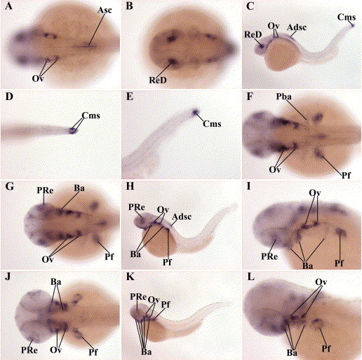
Fig. 6 Distribution of zebrafish crabp2b mRNA during pharyngula stage of embryonic development. Whole mount in situ hybridization showing zebrafish crabp2b mRNA in embryos at 24 hpf (A–E), 36 hpf (F–I) and 48 hpf (J–L) stages. (A, B, D, f, G, I, J, L) Dorsal view, anterior is to left; (C, E, H, K) side view, anterior is to left. The crabp2b mRNA expression in the anterior dorsal spinal cord (Adsc), anterior spinal cord (Asc), branchial arches (Ba), caudal most somite(s) (Cms), otic vesicle (Ov), part of retina (Pre), pectoral fin (Pf), posterior branchial arch (Pba) and retina dorsal to lens (ReD) is indicated.
Reprinted from Gene expression patterns : GEP, 5(3), Sharma, M.K., Saxena, V., Liu, R.Z., Thisse, C., Thisse, B., Denovan-Wright, E.M., and Wright, J.M., Differential expression of the duplicated cellular retinoic acid-binding protein 2 genes (crabp2a and crabp2b) during zebrafish embryonic development, 371-379, Copyright (2005) with permission from Elsevier. Full text @ Gene Expr. Patterns