
Image
|
Figure Caption
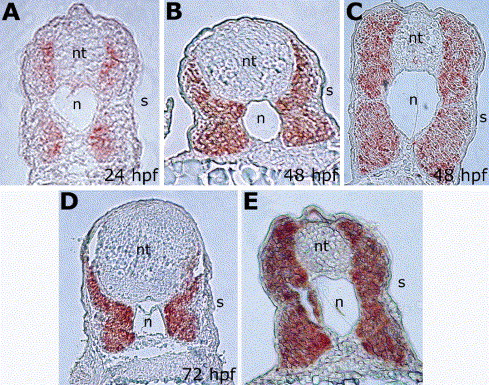
Fig. 6 Zebrafish Grm localization pattern at late stages of development. Localization of Grm protein in the developing somites as shown by immunodetection, on transverse trunk sections at 24 hph (A), 48 hpf (B,C), and 72 hpf (D,E) stages. (D) is the anterior-most section at the hindbrain level, (B) is a section of the anterior spinal cord, and (A,C,E) are posterior trunk sections. Abbreviations: n, notochord; nt, neural tube; s, somites.
Acknowledgments
This image is the copyrighted work of the attributed author or publisher, and
ZFIN has permission only to display this image to its users.
Additional permissions should be obtained from the applicable author or publisher of the image.
Reprinted from Gene expression patterns : GEP, 5(4), Nicoli, S., Gilardelli, C.N., Pozzoli, O., Presta, M., and Cotelli, F., Regulated expression pattern of gremlin during zebrafish development, 539-544, Copyright (2005) with permission from Elsevier. Full text @ Gene Expr. Patterns