
|
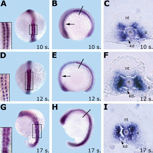
Fig. 4 grm expression pattern during somitogenesis. grm mRNA is detected at 10-somite (A–C), 12-somite (D–F), and 17-somite (G–I) stages, in whole-mounted embryos (A,B,D,E,G,H), and trunk transverse sections (C,F,I). Squares in (A,D,G) are high magnifications of the developing somites (dorsal view), arrows in (B,E) indicate grm expression in the anterior mesoderm, and arrow heads point to the forming longitudinal kidney ducts (C,F,I). Black lines in (B,E,H) indicate the cut position in the correspondent transverse sections (C,F,I). (A,D,G) are dorsal views, anterior to the top, and (B,E,H) are lateral views, anterior to the left. Abbreviations: kd, kidney ducts; n, notochord; nt, neural tube; s, somites.
Reprinted from Gene expression patterns : GEP, 5(4), Nicoli, S., Gilardelli, C.N., Pozzoli, O., Presta, M., and Cotelli, F., Regulated expression pattern of gremlin during zebrafish development, 539-544, Copyright (2005) with permission from Elsevier. Full text @ Gene Expr. Patterns