
Image
|
Figure Caption
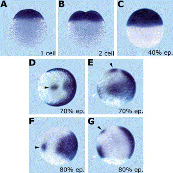
Fig. 3 grm maternal and zygotic expression during early stages of development and gastrulation. Expression pattern of grm as shown by whole-mount in situ hybridization at 1-cell (A), 2-cell (B), 40% epiboly (C), 70% epiboly (D,E), and 80% epiboly (F,G) stages. Black arrow heads point to the grm expression spot on the animal hemisphere (D–G), and white arrow heads in (E,G) indicate grm expression in the ventral mesoderm. (A–C,E,G) are lateral views, (D,F) animal views. Embryos are oriented with dorsal to the right and ventral to the left (D–G).
Figure Data
Acknowledgments
This image is the copyrighted work of the attributed author or publisher, and
ZFIN has permission only to display this image to its users.
Additional permissions should be obtained from the applicable author or publisher of the image.
Reprinted from Gene expression patterns : GEP, 5(4), Nicoli, S., Gilardelli, C.N., Pozzoli, O., Presta, M., and Cotelli, F., Regulated expression pattern of gremlin during zebrafish development, 539-544, Copyright (2005) with permission from Elsevier. Full text @ Gene Expr. Patterns