
|
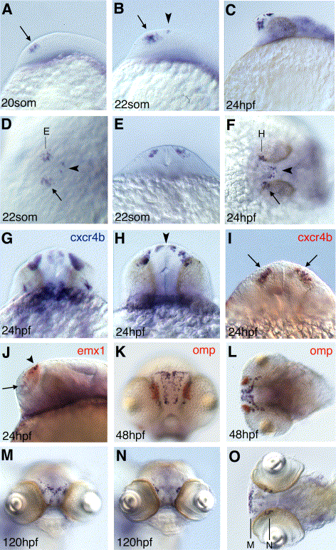
Fig. 2 Neuronal expression of p73. (A–O) In situ hybridization for p73 stained in blue, except (G) which shows staining for cxcr4b. (A–C, J) are lateral views, (D, F, L, O) dorsal views, anterior to the left; (K, M, N) are frontal views; (E, G–I) are optical cross sections, with the focal plane of (E) indicated in (D), focal plane of (H) indicated in (F), and focal planes of (M) and (N) indicated in (O). Developmental stages are indicated in lower left corner; for (O) it is 120 hpf. Probes for double labelings in red are indicated in upper right corner. Arrows in (A, B, D, F, I, J) mark rostro-lateral cell, arrowheads in (B, D, H, J) mark caudo-medial cells.
Reprinted from Gene, 323, Rentzsch, F., Kramer, C., and Hammerschmidt, M., Specific and conserved roles of TAp73 during zebrafish development, 19-30, Copyright (2003) with permission from Elsevier. Full text @ Gene