
|
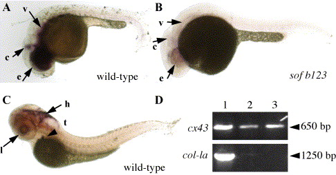
Fig. 3 Expression of connexin 43 in embryos. In situ hybridization was completed using a cx43 probe against the cx43 3′ UTR. (A) Expression of cx43 in wild-type embryo at 24 hpf. e, eyes; c, cerebellum; v, vasculature. (B) Expression of cx43 in sof embryo at 24 hpf. e, eyes; c, cerebellum; v, vasculature. (C) Expression of cx43 in wild-type larvae at 72 hpf. Embryos were treated with PTU to prevent the production of melanin and facilitate the identification of stained structures. l, lens epithelium; h, hindbrain; t and arrowhead, thymus. (D) RT-PCR from 72 hpf whole embryos (lane 1), 72 hpf embryonic hearts (lane 2), and adult hearts (lane 3). The top panel shows amplification from cx43-3′ UTR primers, the bottom panel shows amplification from col-1a primers. Approximate sizes of amplified products are shown on the right (arrowhead).
Reprinted from Developmental Biology, 278(1), Iovine, M.K., Higgins, E.P., Hindes, A., Coblitz, B., and Johnson, S.L., Mutations in connexin43 (GJA1) perturb bone growth in zebrafish fins, 208-219, Copyright (2005) with permission from Elsevier. Full text @ Dev. Biol.