
|
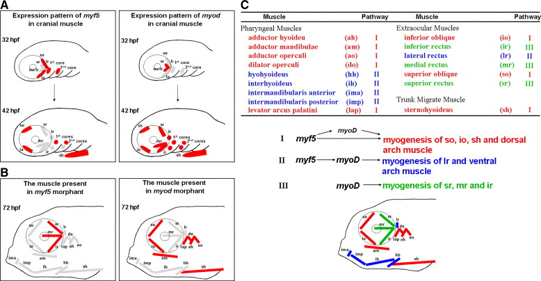
Fig. 9 A plausible model to present the distinct modulation of Myf5 and Myod during craniofacial myogenesis of zebrafish. (A) Schematic illustration of the dynamic expression of myf5 and myod in the cranial muscle of zebrafish embryos at 32 and 42 hpf. The myf5- or myod-positive muscle fibers are labeled in red; myf5- and myod-negative ones are labeled in gray. (B) Schematic illustration of the presence (red) or absence (gray) of cranial muscle fibers in the embryos injected with either myf5-morpholino oligonucleotide (MO; left) or myod-MO (right) at 72 hpf. (C) Schematic diagram of all cranial muscles that are categorized into three groups (represented in red, blue, and green) on the basis of three regulatory pathways of myf5 and myod during development: (I) for so, io, sh, and dorsal arch, not only does myf5 modulate myogenesis directly to generate myogenesis at the basal level but myf5 also triggers myod expression to enhance myogenesis at a high level; (II) for lr and ventral arch, myf5 defines the cell fate of muscle and myod is the major factor of myogenesis; and (III) for sr, mr, and ir, myod modulates myogenesis directly.
Reprinted from Developmental Biology, 299(2), Lin, C.Y., Yung, R.F., Lee, H.C., Chen, W.T., Chen, Y.H., and Tsai, H.J., Myogenic regulatory factors Myf5 and Myod function distinctly during craniofacial myogenesis of zebrafish, 594-608, Copyright (2006) with permission from Elsevier. Full text @ Dev. Biol.