
|
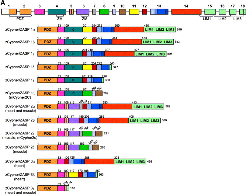
Fig. 1 (A) Genomic organization of cypher and the 13 identified splice variants from zebrafish heart and skeletal muscle. Shown on top is a schematic for the genomic structure of the zebrafish cypher gene which is found on chromosome 13 and spans a region of 80.125 bp. It consists of 18 exons with exon 4 and 6 containing the ZM motifs. Shown below are thirteen different splice variants (all sequences have been submitted to Genbank). The cypher1 splice variants (containing exon 4) were only found in heart tissue, for the other variants, it is indicated whether they have been found in skeletal muscle (labeled “muscle”) or heart or in both tissues. The cypher1ζ form shows similarity to the mouse Cypher2c splice form and cypher2γ shows similarity to the murine Cypher2s. All other splice forms are novel, and similar splice forms from other species have not been described yet. (B) Alignment of zebrafish cypher2γ to human ZASP and mouse Cypher2s and a species comparison of the ZM motif containing exons 4 and 6. Shown on top is the comparison of the amino acid (aa) sequences of the three homologous splice variants from zebrafish (cypher2γ submitted to Genbank), human (ZASP Accession number: AJ133766), and mouse Cypher2s (accession number AF114379). The sequence identities on the aa sequences are: mouse vs zebrafish 70.6%; human vs zebrafish 66.7%; and mouse vs human 92.4%. The exons are marked on top and correspond to the zebrafish gene. Shown in the middle is the alignment of exon 4 found exclusively in heart-specific splice variants. The sequence comparison of exons 4 and 6 is based on the genomic sequences of the different species. The ZM motif is marked on top, and mutations identified in human patients with myopathies are indicated by arrows below. Shown in the bottom is a similar alignment for exon 6 as shown above for exon 4. Arrows indicate mutations described for human patients with myopathies (red arrows refer to mutations described by Vatta et al., 2003 and black arrows indicate mutations described by Selcen and Engel, 2005).
Reprinted from Developmental Biology, 299(2), van der Meer, D.L., Marques, I.J., Leito, J.T., Besser, J., Bakkers, J., Schoonheere, E., and Bagowski, C.P., Zebrafish cypher is important for somite formation and heart development, 356-372, Copyright (2006) with permission from Elsevier. Full text @ Dev. Biol.