
Image
|
Figure Caption
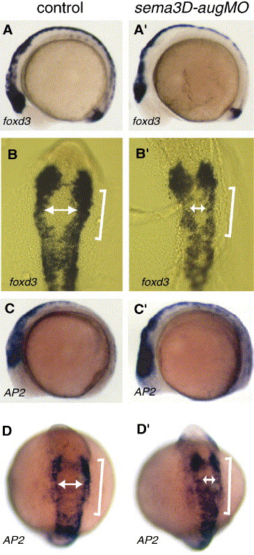
Fig. 4 Migratory defects were observed with foxd3 and AP2 in sema3D-aug morphants. In situ hybridization with foxd3 (A, A′, B, B′) and AP2 (C, C′, D, D′). Both markers were expressed including the cranial neural crest at 8 SS. Dorsal views (B, B′, D, D′, cranial up) showed that cells expressing both markers in morphants did not migrate as far as those in wild type, suggesting that migratory pattern was disrupted. Brackets in panels B, B′, D and D′ showed altered pattern of migration. White arrows in panels B, B′, D and D′ indicated the distance of migrating neural crest cells.
Figure Data
Acknowledgments
This image is the copyrighted work of the attributed author or publisher, and
ZFIN has permission only to display this image to its users.
Additional permissions should be obtained from the applicable author or publisher of the image.
Reprinted from Developmental Biology, 298(1), Sato, M., Tsai, H.J., and Yost, H.J., Semaphorin3D regulates invasion of cardiac neural crest cells into the primary heart field, 12-21, Copyright (2006) with permission from Elsevier. Full text @ Dev. Biol.