
|
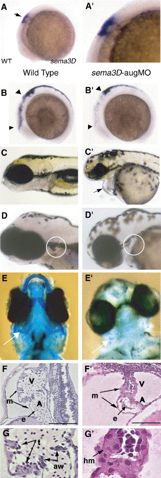
Fig. 2 sema3D-aug morphants have cardiac neural crest defects. (A, A′) At 8 SS, sema3D RNA is expressed in rhombomere 3–5 and in neural crest (black arrow). Anterior left, dorsal up. (B–F′) Phenotypic analyses of wild-type (B–F) and sema3D-augMO morphants (B′–F′). Morphants had reduced crestin expression in cranial crest (8SS, arrowheads, B and B′), pericardiac edema (4 dpf, arrow c′) and shortened neck indicative of pharyngeal arch defects, decreased rag1 expression in the thymus (white circle, D′), and defective facial and pharyngeal cartilage development (white arrow, alcian blue staining, ventral view, E′). These phenotypes are indicative of neural crest defects. (F, F′) Sema3D morphants have dysmorphic hearts with smaller ventricle (V), smaller atrium (A) and thickened myocardium (m). Endocardium was present (e), but AV valve and trabeculation were absent (4 dpf, lateral section, H&E stain, 100 μm scale bar). (G, G′) Magnified pictures of F and F′, respectively. Trabeculation (t) and AV valve formation (avv) observed in wild-type embryo was not found in morphant. In morphant, hypertrophic cardiomyocytes (hm) were observed.
Reprinted from Developmental Biology, 298(1), Sato, M., Tsai, H.J., and Yost, H.J., Semaphorin3D regulates invasion of cardiac neural crest cells into the primary heart field, 12-21, Copyright (2006) with permission from Elsevier. Full text @ Dev. Biol.