
|
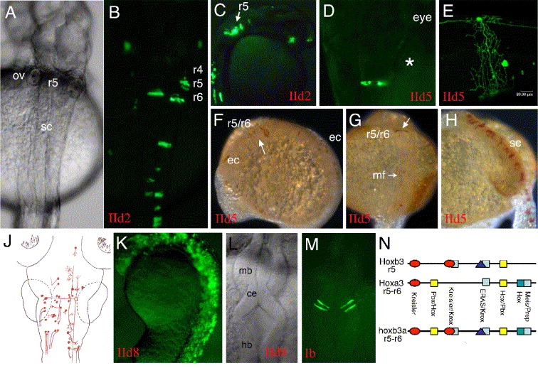
Fig. 8 (A to J) Sequences upstream of hoxb3a exon 4 (part of transcript I) were tested for enhancer activity in combination with the endogenous hoxb3a P1. Construct IId2 included 1.7 kb upstream of exon 4 and contained the CNR15 (Fig. 2). This fragment was capable of directing strong mosaic EGFP expression within the spinal cord and hindbrain up to the r4/r5 boundary. When CNR14 was included into the construct (IId5), the same expression pattern was observed. Labeled embryos shown in pictures A to J are representative for the types of EGFP expressing neurons that were observed with both constructs. (A, B) Embryo 30 hpf, dorsal view. (C) Embryo 27 hpf, sagittal view. (D) EGFP-labeled neuron in r7 of a larva at 2 dpf. (E) EGFP-labeled spinal cord neuron with its axons surrounding and innervating the yolk. (F–H) IId5-injected embryos stained by anti-GFP immunohistochemistry. (F) 10 somite stage (14 hfp) embryo with EGFP expression in clonal strings within r5 and r6 and posterior. (G) 20 somite stage (19 hpf) embryo with r5 and r6 and muscle fiber label. (H) 24 hpf embryo displaying strong label in clonal strings within the spinal cord. (J) Illustration of the types of neurons that were EGFP labeled. (K) CNR14 was analyzed together with a CNR15/CNR16 fragment (IId8; Fig. 2). (L and M) Activity of the mouse CNR15 enhancer in clonal strings within the anlage of the cerebellum is shown. This enhancer mediated activity within the central nervous system, but was not able to set an anterior expression boundary within the hindbrain. (N) Schematic illustration of the structural similarities of zebrafish hoxb3a r5–r6 enhancer (CNR15) with mouse Hoxa3 r5–r6 and mouse Hoxb3 r5 enhancer sequences (compare Manzanares et al., 1999 and Hadrys et al., 2004). Abbreviations: ce, cerebellum; ec, epithelial cells; hb, hindbrain; mb, midbrain; ov, otic vesicle; r, rhombomere; sc, spinal cord.
Reprinted from Developmental Biology, 297(1), Hadrys, T., Punnamoottil, B., Pieper, M., Kikuta, H., Pezeron, G., Becker, T.S., Prince, V., Baker, R., and Rinkwitz, S., Conserved co-regulation and promoter sharing of hoxb3a and hoxb4a in zebrafish, 26-43, Copyright (2006) with permission from Elsevier. Full text @ Dev. Biol.