
|
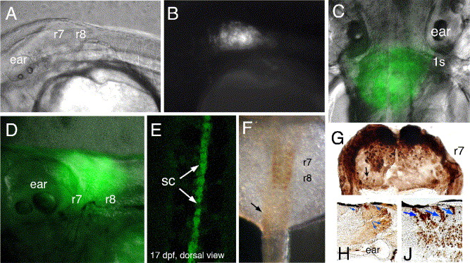
Fig. 7 Zebrafish line CLGY838 with viral-YFP insertion 870 bp upstream of CNR10 (compare Fig. 2). Expression of YFP becomes progressively stronger during zebrafish development as shown here (A, B) for 32 hpf embryos, (C) 4 dpf, and (D) 10 dpf larvae. YFP expression domain includes r7 and r8 extending into the posterior r6, here forming a diffuse boundary. (E) Spinal cord expression in 17-dpf larva from dorsal view. (F) Antibody stained 32 hpf embryo reveals r7 and r8 expression and low YFP levels within the somitic mesoderm. (G) Coronal section through hindbrain r7 of 4-day-old larva stained with an antibody against GFP. YFP expressing cells are localized in the dorsomedial region, note also single neurons that are stained in the ventral area (arrows). (H and J) Sagittal sections through 26 dpf larva stained by anti-GFP immunohistochemistry. (H) Section through the lateral hindbrain. Blue arrows point to neuronal cell clusters and single cells. (J) Section through the medial hindbrain. Hoxb4a expressing cells in the dorsomedial hindbrain form a stripe pattern (blue arrows) suggesting streams of migrating cells. Abbreviations: r, rhombomere; s, somite; sc, spinal cord.
Reprinted from Developmental Biology, 297(1), Hadrys, T., Punnamoottil, B., Pieper, M., Kikuta, H., Pezeron, G., Becker, T.S., Prince, V., Baker, R., and Rinkwitz, S., Conserved co-regulation and promoter sharing of hoxb3a and hoxb4a in zebrafish, 26-43, Copyright (2006) with permission from Elsevier. Full text @ Dev. Biol.