
|
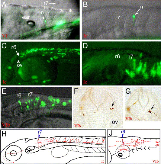
Fig. 6 The regulatory function of the genome region that includes CNR10 and CNR11 was investigated. (A) CNR11 in the context of hoxb4a P1 (construct Vf; Fig. 2) repressed the broad activity of the hoxb4a P1 fragment and enhanced r7 and r8 EGFP expression. Clear neural expression was first detected at 24 hpf in single neurons within hindbrain r7, r8, and spinal cord. A 3-dpf larva is shown that has EGFP label in r7 and r8 as well as in single muscle fibers. (B) CNR11 acts also as a neural enhancer in r7 and r8 when combined with hoxb3a/hoxb4a P2 (CNR1) that alone mediates only very weak transcriptional activity within the spinal cord (compare Fig. 3A, Table 3). (C and D) A 2.3-kb genome fragment that included CNR10 and CNR11 (Fig. 2) in combination with hoxb3a/hoxb4a P2 (CNR1) drove strong EGFP expression up to the r6/r7 boundary within r7 and r8 and within the spinal cord. Also muscle fibers were labeled. (E) When CNR1 was excluded from the construct and the hoxb3a transcript III promoter sequence that is included in CNR11 (hoxb3a P3) was used to drive Gal4 expression, the anterior EGFP expression boundary was moved anteriorly. EGFP-labeled neurons were observed in r5 and r6, but never in r4. (F and G) 30-μm vibratome sections through embryos that expressed the CNR10/11 reporter gene construct and were stained with an anti GFP antibody, on level of former r5 (F), and on former r7 level (G). (H) Schematic illustration of the types of neurons that were observed by injection of the hoxb3a/hoxb4a P2-CNR10/CNR11 reporter construct (Ic). (J) Types of neurons that were observed with the CNR10/CNR11 (hoxb3a P3) construct (VIb). Abbreviations: n, neuron; ov, otic vesicle; r, rhombomere; s, somite.
Reprinted from Developmental Biology, 297(1), Hadrys, T., Punnamoottil, B., Pieper, M., Kikuta, H., Pezeron, G., Becker, T.S., Prince, V., Baker, R., and Rinkwitz, S., Conserved co-regulation and promoter sharing of hoxb3a and hoxb4a in zebrafish, 26-43, Copyright (2006) with permission from Elsevier. Full text @ Dev. Biol.