
|
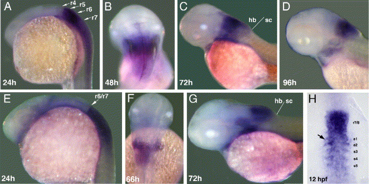
Fig. 4 Whole mount in situ hybridization with hoxb3a exon 6 (A–D) and hoxb4a exon 3 (E–H) probe. (A) Hoxb3a transcript distribution in r5 and r6 extending posterior into the spinal cord in 24 hpf embryos. (B–D) Gene expression proceeds within the hindbrain, strongest on former r4 to r6 level, in 48 hpf (B), 72 hpf (C), and 96 hpf (D) larvae. (E) Hoxb4a is expressed within the spinal cord and in r7 and r8 with a sharp border at the boundary between r6 and r7 in embryos at 24 hpf. (F and G) Expression domain in later development becomes increasingly confined to the posterior hindbrain in 66 hpf (F) and 72 hpf (G) larvae. (H) Somitic expression of hoxb4a in early embryos. Abbreviations: hb, hindbrain; r, rhombomere; sc, spinal cord.
Reprinted from Developmental Biology, 297(1), Hadrys, T., Punnamoottil, B., Pieper, M., Kikuta, H., Pezeron, G., Becker, T.S., Prince, V., Baker, R., and Rinkwitz, S., Conserved co-regulation and promoter sharing of hoxb3a and hoxb4a in zebrafish, 26-43, Copyright (2006) with permission from Elsevier. Full text @ Dev. Biol.