
|
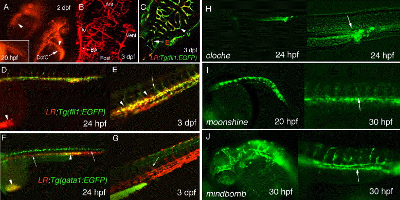
Fig. 2 The lmo2 transgenic lines facilitate visualization of hematopoietic and vascular systems in wild-type and mutant embryos. (A) In LR embryos, DsRed protein is initially detected at 20 hpf (inset); 2 dpf LR embryos labeling hematopoietic (white arrowheads) and endothelial cells (DofC: ducts of Cuvier). (B) The cranial vessels of a 3 dpf LR embryo visualized by confocal microscopy (BA, basilar artery; Ant, Anterior; Post, posterior; Do, dorsal; Vent, ventral). (C) Confocal section through the head and trunk of a 3 dpf LR; Tg(fli1:EGFP) embryo displaying green/red cranial vessels (arrow, V) and red erythrocytes (arrowhead, E). (D,E) LR; Tg(fli1:EGFP) embryos distinctly label hematopoietic (arrowhead) and endothelial cells (arrows) in 24 hpf and 3 dpf embryos. In LR; Tg(gata1:EGFP) transgenic embryos, green/red erythrocytes (arrowheads) circulate through vessels (arrows) labeled by DsRed as shown in 24 hpf and 3 dpf embryos (F and G, respectively). In the second panel, 24–36 hpf mutant embryos carry the Tg(lmo2:EGFP) transgene. (H) In the zebrafish cloche mutant, there is an absence of hematopoietic cells and virtually no endothelium; EGFP expression is restricted to the posterior ICM (arrow), where lmo2 expression is known to be preserved. (I) In moonshine, normal vasculature is present (arrow), but erythrocytes are absent. (J) In mindbomb, vascular malformations (arrow points to fusion between artery and vein) are identified when EGFP-labeled cells travel from aorta into vein.
Reprinted from Developmental Biology, 281(2), Zhu, H., Traver, D., Davidson, A.J., Dibiase, A., Thisse, C., Thisse, B., Nimer, S., and Zon, L.I., Regulation of the lmo2 promoter during hematopoietic and vascular development in zebrafish, 256-269, Copyright (2005) with permission from Elsevier. Full text @ Dev. Biol.