
|
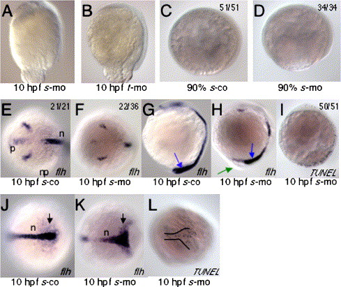
Fig. 4 sid4 morpholinos cause morphogenetic defects and increased apoptosis. (A) s-mo and (B) t-mo produce similar epiboly defects. In these embryos, the blastoderm margin appears to constrict before the blastoderm completely covers the yolk. (C) Actively gastrulating control and (D) morphant embryos (90% epiboly) do not exhibit apoptosis. (E) Flh is appropriately expressed in the neural plate and polster of 10 hpf s-co and (F) s-mo embryos. The number of anterior flh+cells appears reduced in the morphants. (G) Flh-expressing cells extend into the tail bud (blue arrow) of s-co embryos. (H) Yolk extrusions (green arrow) inhibit normal tail bud formation and patterning of caudal flh+cells. (I) Apoptosis increases in the caudal mesoderm of 10 hpf morphants. (J) Caudal flh-expressing cells (black arrows) converge normally and extend to form a narrow, compact notochord. (K) Convergence is impaired in morphant embryos and the notochord field is much broader than controls. (L) In many cases, the distribution of apoptotic cells in 10 hpf morphants was similar to flh expression patterns. Ratios of embryos exhibiting a given phenotype are in upper right. Lateral views in A–D; G–I. Dorsal views in E, F, J–L. Embryonic stages and treatments are indicated below each panel. In situ probes and TUNEL are indicated in lower right.
Reprinted from Developmental Biology, 282(1), diIorio, P.J., Runko, A., Farrell, C.A., and Roy, N., Sid4: A secreted vertebrate immunoglobulin protein with roles in zebrafish embryogenesis, 55-69, Copyright (2005) with permission from Elsevier. Full text @ Dev. Biol.