
Fig. 7
- ID
- ZDB-IMAGE-070925-92
- Genes
- Publication
- Gulati-Leekha et al., 2006 - A reporter-assisted mutagenesis screen using α1-tubulin-GFP transgenic zebrafish uncovers missteps during neuronal development and axonogenesis
- All Figures
- Figures for Gulati-Leekha et al., 2006
|
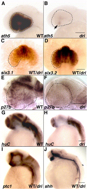
Fig. 7 Mutation in drishti causes an early cell cycle block, which portends the later neurogenesis defects. ISH carried out in phenotypically normal embryos, 25% of which show visibly reduced ath5 expression except in the ventronasal patch (arrow; panels A and B). Prior to this defect, six3 regulation is unperturbed at the 15-somite stage (C, D). Panels E and F highlight an accompanying decrease in the cell cycle marker, p27b, at 34 hpf. At the same time, fewer postmitotic Hu-positive neurons are observed in the developing CNS (G, H). Expression of patched1 (ptc1), an indicator of Hh signaling as well as sonic hedgehog (shh), is indistinguishable in a pool of 34 hpf embryos (I, J). All images show lateral views of embryos with anterior towards left except C and D (dorsal views with anterior towards top). Scale bars: 100 μm in all panels.
Reprinted from Developmental Biology, 296(1), Gulati-Leekha, A., and Goldman, D., A reporter-assisted mutagenesis screen using α1-tubulin-GFP transgenic zebrafish uncovers missteps during neuronal development and axonogenesis, 29-47, Copyright (2006) with permission from Elsevier. Full text @ Dev. Biol.