
Fig. 3
- ID
- ZDB-IMAGE-070925-84
- Genes
- Publication
- Gulati-Leekha et al., 2006 - A reporter-assisted mutagenesis screen using α1-tubulin-GFP transgenic zebrafish uncovers missteps during neuronal development and axonogenesis
- All Figures
- Figures for Gulati-Leekha et al., 2006
|
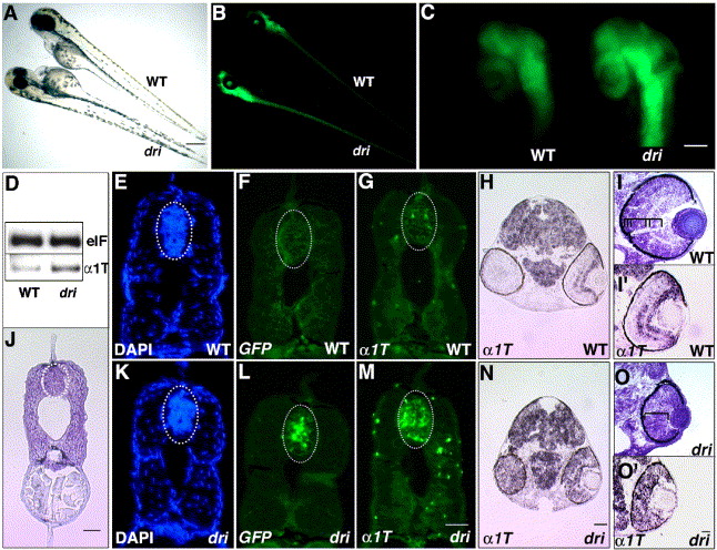
Fig. 3 Mutation in drishti increases GFP and α1-tubulin mRNA in the developing CNS. Bright-field (A) and fluorescent (B) images of dri mutant and a sibling at 3 dpf. Note the panneurally increased GFP expression and reduced eye size. Mutant retinas display a significant increase in fluorescence, clearly visible in PTU-treated embryos (C). RT-PCR at 2 dpf confirms a concomitant increase in α1-tubulin (α1T) mRNA, e-1F4α (e1F) is the normalizing control (D). Transverse sections of 2-day-old embryos show that the dri spinal cord has slightly reduced cell number, 107 ± 8, n = 3, in wild-type and 83 ± 11.3, n = 3, in mutants (DAPI staining, E and K); however, ISH analysis using a GFP (fluorescein-labeled) riboprobe (F and L) and α1-tubulin (fluorescein-labeled) riboprobe (G and M) demonstrate that these transcripts are remarkably higher in mutants (panel J shows a cresyl violet-stained representative section for the spinal cord level at which above analysis was performed). A similar increase in mRNA expression is observed in dri brain at 2 dpf (panels H and N, α1-tubulin ISH using a digoxigenin-labeled riboprobe; GFP ISH not shown). dri retina at 2 dpf lacks the typical laminar organization of its wild-type counterpart (cresyl violet staining, panels I and O) and expresses α1-tubulin at a uniformly high level (digoxigenin-labeled α1T riboprobe, panels I′ and O′) whereas the strongest expression in WT retina is restricted to the GCL (and central INL). Ellipses (E–G, J–M) outline the spinal cord region in which total cell counts were performed. Scale bars: 250 μm in panels A and B; 100 μm in panel C; and 25 μm in panels J, M (for E–G and K–M), N (for H,N) and O′ (for I, I′, O and O′).
Reprinted from Developmental Biology, 296(1), Gulati-Leekha, A., and Goldman, D., A reporter-assisted mutagenesis screen using α1-tubulin-GFP transgenic zebrafish uncovers missteps during neuronal development and axonogenesis, 29-47, Copyright (2006) with permission from Elsevier. Full text @ Dev. Biol.