
|
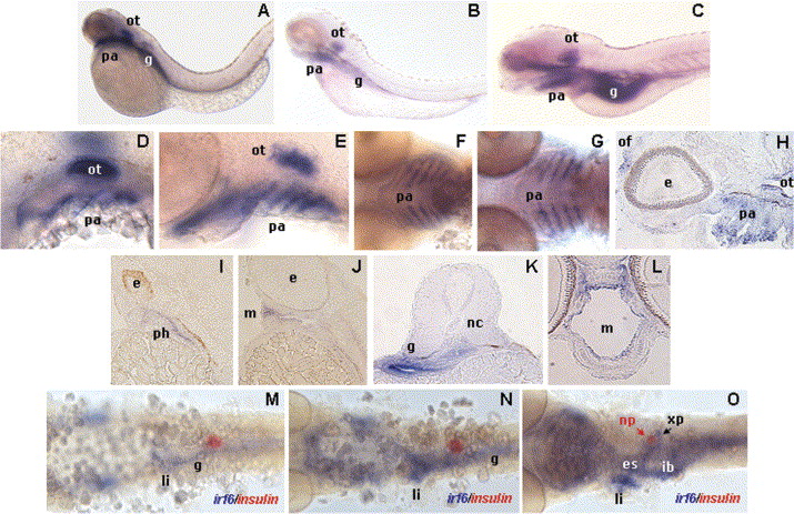
Fig. 4 Expression of irf6 transcript in the developing zebrafish during pharyngula, hatching and larval stages. Whole-mounts of 2 dpf (A) and 3 dpf (B) embryos show strong irf6 expression in the mouth and pharyngeal arch region, with increased expression intensity in 5 dpf larvae (C). Flat-mount lateral (D,E) and ventral (F,G) views, and sagittal section view (H), shows expression in pharyngeal arches at 35 hpf (D), 2 dpf (E), 3 dpf (F), 4 dpf (G), and 5 dpf (H). Sagittal (I,J) and transverse (K,L) tissue sections of stained 2 dpf (I,K), 3 dpf (J), and 5 dpf (H,L) embryos and larvae show localization of irf6 expression in the mouth, pharynx, and gut. Flat-mount dorsal views of digestive system at 35 hpf (M), 2 dpf (N), and 3 dpf (O) reveal irf6 expression in the liver and exocrine pancreas in addition to the esophagus and intestinal bulb (gut). e, Eye; es, esophagus; g, gut; li, liver; m, mouth; nc, notochord; of, olfactory vesicle/naris; ot, otic vesicle; pa, pharyngeal arch; ph, pharynx; np, endocrine pancreas; xp, exocrine pancreas.
Reprinted from Gene expression patterns : GEP, 5(5), Ben, J., Jabs, E.W., and Chong, S.S., Genomic, cDNA and embryonic expression analysis of zebrafish IRF6, the gene mutated in the human oral clefting disorders Van der Woude and popliteal pterygium syndromes, 629-638, Copyright (2005) with permission from Elsevier. Full text @ Gene Expr. Patterns