
|
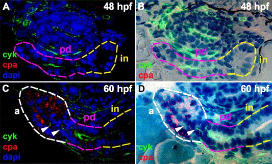
Fig. 3 Exocrine differentiation and gland morphogenesis. (A–D) Histological cross-sections of 48 hpf (A) and 60 hpf (C) larvae processed for whole mount cpa and cyk immunohistochemistry (IHC) and dapi. (B and D) Merged fluorescent images without dapi staining (A and C) and bright field images of the corresponding histological sections stained with methylene blue and azure II. The main pancreatic duct (pink outline) is contiguous with the intestine (in, yellow outline) at 48 hpf. Cpa expression is evident in exocrine cells forming a primitive acinus-like structure at 60 hpf (white outline). Cyk+ duct cells (arrowheads) are present adjacent to the developing acinus but are not contiguous with the extrapancreatic duct in this and adjacent sections (not shown). cpa: carboxypeptidase A; cyk: cytokeratin; pd: pancreatic duct; a: acinus; in: intestine.
Reprinted from Developmental Biology, 284(1), Yee, N.S., Lorent, K., and Pack, M., Exocrine pancreas development in zebrafish, 84-101, Copyright (2005) with permission from Elsevier. Full text @ Dev. Biol.