
|
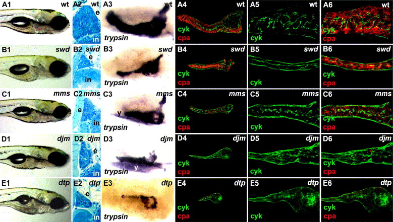
Fig. 9 Mutations affecting late stages of exocrine pancreas development. (A1–E1) Right lateral view of live 5 dpf wild type and mutant larvae. (A1) The wild type intestinal epithelium is folded, a widely patent gut lumen is obvious, and there is little residual yolk. The intestine is grossly normal in swd (B1), mms (C1), djm (D1) and dtp (E1) mutants. In swd, skin pigmentation (melanophores) is reduced, but xanthophores and iridophores appear unaffected (B1). There is a small amount of residual yolk in dtp (E1). (A2–E2) Histological cross-sections through the 5 dpf pancreas, 3 μm caudal to the islet. In wild type (A2), there are multiple exocrine acini; in swd (B2), there are relatively few exocrine acini, and electron micrograph shows that the acinar cells contain zymogen granules (Supplementary Fig. 3D); in mms (C2), multiple acini are observed, but the cytoplasm of the acinar cells contains vacuole-like structures and very few zymogen granules (Supplementary Fig. 3H); in djm (D2) and dtp (E2), there are no recognizable acini present, and the exocrine cells appear undifferentiated. (A3–E3) Right lateral view of whole mount trypsin in situ hybridization on 5 dpf. Strong trypsin expression is evident in the wild type pancreas (A3), whereas in swd (B3), mms (C3), djm (D3) and dtp (E3), trypsin expression is reduced. There is background staining in the yolk. (A4–E4) Visualization of pancreatic ducts and acinar cells by cyk and cpa immunohistochemistry, respectively. The wild type ductal system is composed of a branching network of small ducts and cpa+ acinar cells (A4). The swd pancreas (B4) is small, but there is relatively normal cpa; in the mms pancreas (C4), there are only low levels of cpa. There is no significant cpa detected in djm (D4) and dtp (E4). (A5–E5 and A6–E6) Confocal projection of pancreatic ducts (green) and acini (red) in larvae processed for cyk and cpa immunohistochemistry. Wild type pancreatic ducts are highly branched, and individual acini are each drained by single intercalated duct (A5, 6). In swd (B5, 6), the main pancreatic duct with few branches is evident, whereas in mms (C5, 6), duct branching is evident, albeit less pronounced than in wild type. By comparison, the ductal networks of djm (D5, 6) and dtp (E5, 6) are markedly aberrant. swd: sweetbread; mms: mitomess; djm: ductjam; dtp: ducttrip; e: exocrine pancreas; in: intestine; y: yolk; cyk: cytokeratin; cpa: carboxypeptidase A.
Reprinted from Developmental Biology, 284(1), Yee, N.S., Lorent, K., and Pack, M., Exocrine pancreas development in zebrafish, 84-101, Copyright (2005) with permission from Elsevier. Full text @ Dev. Biol.