
|
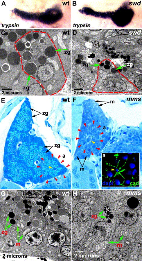
Fig. S3 Exocrine pancreas in the sweetbread (swd) and mitomess (mms) mutants. (A, B) Right lateral view of 72 hpf wild type (A) and swd (B) larvae processed for trypsin whole mount in situ hybridization. There is normal trypsin expression in the 72 hpf swd mutant identified by reduced skin pigmentation (B). (C, D) Acinar cell hypoplasia is evident in 5 dpf swd mutant. Acinar cells (red dashed lines) in swd mutant (D) appear small with relatively few and small zymogen granules (indicated by green arrows), compared with wt sibling (C). (E, F) Histological cross-sections through the pancreatic tail of 5 dpf wild type (E) and mms (F) mutant larvae stained with methylene blue and azure II. In routine histological sections, acinar gland morphology is difficult to appreciate. However, both in wild type and mms mutants, acinar cells surrounding centroacinar cells (yellow arrowheads) can be discerned. Red arrow heads outline acinar borders. Note the zymogen granules are prominent in wild type and they are relatively few and small in mms. Also note the cytoplasmic vacuole-like structures in mms acinar cells. Electron microscopy identifies these as swollen mitochondria (below). Inset (a) shows preserved mms acinar gland morphology as revealed by cadherin (green) immunohistochemistry and dapi (blue). (G, H) Transmission electron micrographs of 5 dpf wild type and mms acinar cells. Note reduced numbers and size of mms acinar zymogen granules and enlarged swollen mitochondria. zg: zymogen granules; m: mitochondria; a: acinus; cad: cadherin.
Reprinted from Developmental Biology, 284(1), Yee, N.S., Lorent, K., and Pack, M., Exocrine pancreas development in zebrafish, 84-101, Copyright (2005) with permission from Elsevier. Full text @ Dev. Biol.