
|
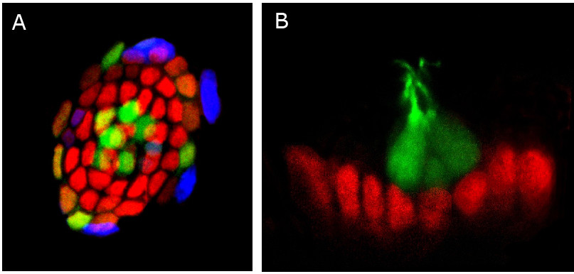
Fig. 5 Distribution of Sox2 in lateral line neuromasts. (a) Confocal image of a zebrafish lateral line neuromast. Mantle cells and hair cells are labeled by GFP (green) in this transgenic line of zebrafish. Fluorescent immunostaining reveals expression of the neural progenitor marker protein Sox2 (red) and BrdU incorporation (blue). Cell division is occurring mostly in the periphery of the neuromast. (b) Confocal image of a zebrafish lateral line neuromast inmunostained to detect the neural progenitor marker protein Sox2 (red) and GFP (green) in hair cells. Note that these two markers do not overlap, suggesting that Sox2 is not present in differentiated cell types in neuromasts. Pictures provided by M Allende.