
|
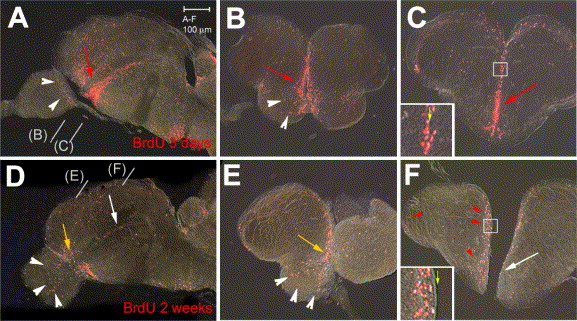
Fig. 3 BrdU-labeled cells (red staining) in the zebrafish adult telencephalon 3 days (A–C) or 2 weeks (D–F) after BrdU injection. All views are vibratome sections observed under conventional fluorescence microscopy (A,D: parasagittal sections, anterior left; B, C, E, F: cross-sections at the levels indicated in panels A, D, dorsal up). Note major changes after 2 weeks: (i) the ventral subpallial stripe is diminished caudally (white arrows in panels D, F) and mostly maintained anteriorly (yellow arrows in panels D, E), (ii) labeled cells are observed in the OB (white arrowheads in panels D, E), and (iii) labeled cells have left the ventricle in the pallium and dorsal subpallium (red arrowheads in panel F, compare insets in panels panels C, F, yellow arrow to the ventricle).
Reprinted from Developmental Biology, 295(1), Adolf, B., Chapouton, P., Lam, C.S., Topp, S., Tannhauser, B., Strähle, U., Gotz, M., and Bally-Cuif, L., Conserved and acquired features of adult neurogenesis in the zebrafish telencephalon, 278-293, Copyright (2006) with permission from Elsevier. Full text @ Dev. Biol.