
|
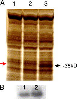
Fig. 8 Sid4 protein synthesis in embryos injected with mRNA and DNA. (A) Protein extracts from 12 hpf uninjected controls (lane 1), and those injected with 500 pg/nl sid4 mRNA (lane 2) exhibit similar amounts of Sid4 protein (red arrow). Robust Sid4 protein synthesis can be seen in extracts from embryos injected with 25 pg/nl pCS2sid4 DNA (Lane 3, black arrow). This new protein migrates with a mass of ∼38 kDa, the predicted size of Sid4 after signal peptide cleavage. (B) In vitro synthesized sid4 mRNA retains its integrity during microinjection. Approximately 2 µg of pre-injection (lane 1) and post-injection (lane 2) mRNA was separated on standard 0.8% agarose gels and stained with ethidium bromide.
Reprinted from Developmental Biology, 282(1), diIorio, P.J., Runko, A., Farrell, C.A., and Roy, N., Sid4: A secreted vertebrate immunoglobulin protein with roles in zebrafish embryogenesis, 55-69, Copyright (2005) with permission from Elsevier. Full text @ Dev. Biol.