
Image
|
Figure Caption
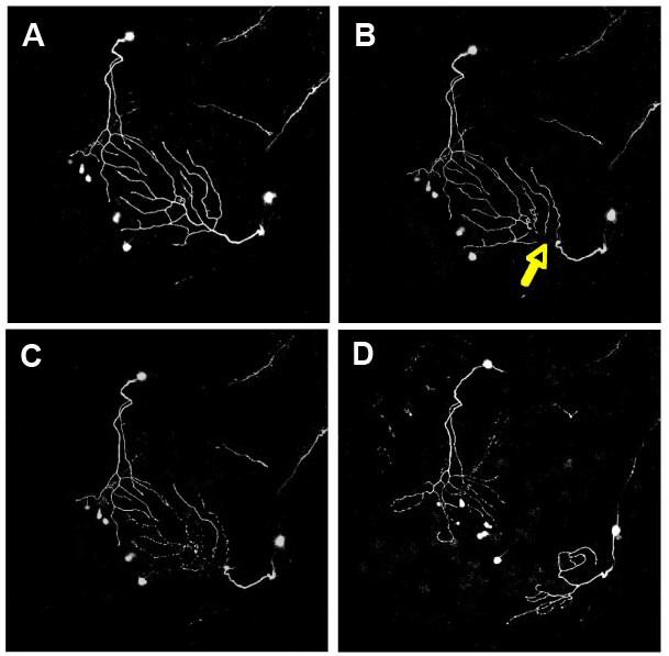
Fig. 3 Imaging peripheral arbor re-innervation in vivo. Confocal projections showing dorsal views of two trigeminal axons, visualized with GFP, in a 54 hpf zebrafish embryo at several time points during a two-photon axotomy experiment. Anterior is to the bottom left. Images are each 420 microns across. (a) Twenty minutes before axotomy. (b) Approximately 20 minutes after axotomy. Yellow arrow points to site of axotomy. (c) Two hours after axotomy, the distal portion degenerates. (d) Robust regrowth is apparent 12 hours after axotomy. Figure provided by A Sagasti.
Acknowledgments
This image is the copyrighted work of the attributed author or publisher, and
ZFIN has permission only to display this image to its users.
Additional permissions should be obtained from the applicable author or publisher of the image.
Full text @ Neural Dev.