
Image
|
Figure Caption
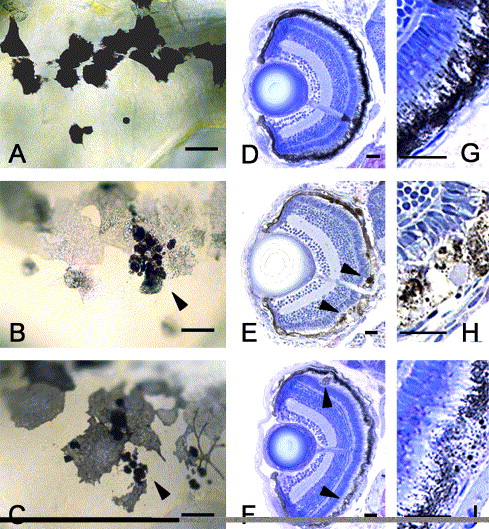
Fig. 6 Phenocopy of fdv by silva knock-down. (C, F, I) Morpholino-oligonucleotide-injected 5-day-old larvae show the same phenotype as (B, E, H) 5-day-old fdv mutant larvae. Compared to the (A, D, G) wild-type, the melanocytes are lighter and contain pigment clots (arrowheads in panels C and F). In the sections taken from (arrowheads in panel F) larvae after morpholino injection, some RPE cells are bloated and contain vacuoles as can be seen in (arrowheads in panel E) fdv mutant larvae. Scale bars in panels A–C are 20 μm, in panels D–F 100 μm and in panels G–I 50 μm.
Acknowledgments
This image is the copyrighted work of the attributed author or publisher, and
ZFIN has permission only to display this image to its users.
Additional permissions should be obtained from the applicable author or publisher of the image.
Reprinted from Developmental Biology, 284(2), Schonthaler, H.B., Lampert, J.M., von Lintig, J., Schwarz, H., Geisler, R., and Neuhauss, S.C., A mutation in the silver gene leads to defects in melanosome biogenesis and alterations in the visual system in the zebrafish mutant fading vision, 421-436, Copyright (2005) with permission from Elsevier. Full text @ Dev. Biol.