
|
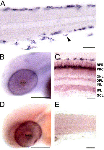
Fig. 5 Spatio-temporal expression pattern of silva and silvb. Expression analyses in 2-day-old wild-type larva. (A and B) silva is expressed in (arrowheads in A) melanocytes and (B) the RPE of the eye. (C) On adult sections, the silva transcript is confined to cells of the RPE and to the melanocytes located behind the eye. (D and E) silvb expression is restricted to RPE cells. RPE, retinal pigment epithelium, PRC, photoreceptor outer segments, ONL, outer nuclear layer, OPL, outer plexiform layer, INL, inner nuclear layer, IPL, inner plexiform layer, GCL, ganglion cell layer. Scale bar in panel A is 50 µm, in panels B and D 100 μm, in panel C 20 μm and in panel E 50 μm.
Reprinted from Developmental Biology, 284(2), Schonthaler, H.B., Lampert, J.M., von Lintig, J., Schwarz, H., Geisler, R., and Neuhauss, S.C., A mutation in the silver gene leads to defects in melanosome biogenesis and alterations in the visual system in the zebrafish mutant fading vision, 421-436, Copyright (2005) with permission from Elsevier. Full text @ Dev. Biol.